Bismuth vs. Mercury Electrodes for Lead Detection: A Comprehensive Performance Comparison for Researchers
This article provides a critical analysis for researchers and scientists on the performance of bismuth-based electrodes as an environmentally friendly alternative to traditional mercury electrodes for the electrochemical detection of...
Bismuth vs. Mercury Electrodes for Lead Detection: A Comprehensive Performance Comparison for Researchers
Abstract
This article provides a critical analysis for researchers and scientists on the performance of bismuth-based electrodes as an environmentally friendly alternative to traditional mercury electrodes for the electrochemical detection of lead. Covering foundational principles, modern sensor designs, and optimization strategies, it delivers a rigorous, evidence-based comparison of sensitivity, selectivity, and practical applicability in complex matrices. The review synthesizes recent advancements to guide material selection and method development, addressing key challenges and future directions for integrating these sensors into biomedical and environmental monitoring applications.
Foundations of Lead Detection: Mercury's Legacy and the Rise of Bismuth
For decades, mercury-based electrodes were the cornerstone of electrochemical stripping analysis for trace heavy metals. Their widespread adoption was fueled by a combination of exceptional analytical properties, including a wide cathodic potential window and highly reproducible surfaces [1]. The inherent toxicity of mercury, however, has driven the scientific community to seek safer, high-performing alternatives [2]. Among these, bismuth-film electrodes have emerged as the most promising successor. This guide provides an objective comparison of the performance between traditional mercury and modern bismuth-based electrodes, with a specific focus on lead detection, contextualized within the broader thesis of evolving electrochemical sensor technology.
Performance Comparison: Bismuth vs. Mercury Electrodes for Lead Detection
The transition from mercury to bismuth electrodes is supported by extensive experimental data. The following tables summarize key performance metrics from recent research, enabling a direct comparison of their analytical capabilities for detecting lead (Pb) and other heavy metals.
Table 1: Direct Performance Comparison of Mercury and Bismuth Film Electrodes for Lead Detection
| Electrode Type | Detection Limit for Pb (µg/L) | Linear Range (µg/L) | Reproducibility (RSD % for Pb) | Key Advantages | Reported Drawbacks |
|---|---|---|---|---|---|
| Mercury Film Electrode (MFE) | 0.1 [1] | 0.1 - 10,000 [1] | ~2.4% [2] | Wide cathodic window, high reproducibility, well-established methodology [1]. | High toxicity, occupational health risks, potential future regulatory restrictions [1] [2]. |
| Nafion-Coated Bismuth Film Electrode (NCBFE) | 0.17 [2] | Not Specified | 2.4% [2] | Low toxicity, insensitive to dissolved oxygen, "environmentally friendly" [2]. | Performance can be dependent on film preparation method. |
| Bismuth Film on GCE (in situ) | 0.93 [3] | Not Specified | High accuracy and repeatability [3] | Effective Hg-alternative, forms alloys with heavy metals, good sensitivity [3]. | -- |
| Bi/DL-Ti3C2Tx/GCE Nanocomposite | 1.73 [4] | 1 - 250 [4] | Good repeatability and stability [4] | Enhanced sensitivity from nanomaterials, low toxicity, high conductivity [4]. | More complex electrode preparation. |
Table 2: Performance in Simultaneous Metal Detection and Real Sample Analysis
| Electrode Type | Simultaneously Detected Metals | Performance in Real Samples | Sample Matrix |
|---|---|---|---|
| Mercury Film Electrode (MFE) | Cd(II), Pb(II), In(III), Cu(II) [1] | Accurate determination of metals in tap water with standard addition methodology [1]. | Tap Water |
| Nafion-Coated Bismuth Film Electrode (NCBFE) | Pb(II), Cd(II), Zn(II) [2] | Results for Pb and Cd in vegetable samples were in agreement with GFAAS [2]. | Vegetable Samples |
| Bismuth Film on GCE | Zn, Cd, Pb, Cu [3] | High accuracy and repeatability within measurements [3]. | Not Specified |
| Bi/DL-Ti3C2Tx/GCE Nanocomposite | Pb(II), Cd(II) [4] | Easy, sensitive, and selective application in actual water samples [4]. | Actual Water Samples |
Experimental Protocols for Electrode Preparation and Analysis
To ensure reproducibility and facilitate comparison, the standard protocols for preparing and utilizing these electrodes are detailed below.
Fabrication of Paper-Based Carbon Electrodes (Substrate)
A common low-cost substrate used in modern studies involves paper-based working electrodes [1].
- Patterning: Hydrophobic wax barriers are printed on chromatography paper (e.g., Whatman Grade 1) using a wax printer.
- Melting: The paper is heated to 80°C to melt the wax, which penetrates the paper to form hydrophobic-hydrophilic patterns.
- Carbon Coating: A carbon ink suspension is drop-cast (e.g., 2 µL) onto one side ("bottom side") of the paper to create the conductive working electrode surface [1].
Mercury vs. Bismuth Film Modification
The modification of the carbon substrate is a critical step that defines the sensor's performance.
Mercury Film Deposition: A thin mercury film is electrochemically deposited onto the carbon surface from a solution of mercury(II) acetate in 0.1 M HCl by applying a negative potential. This significantly reduces the amount of mercury used compared to traditional mercury electrodes [1].
Bismuth Film Deposition (In-Situ Method): This is the most common approach for bismuth film preparation. A solution containing the target analytes (e.g., Pb²⁺, Cd²⁺) also contains Bi³⁺ ions (e.g., 400 µg/L). During the preconcentration step, both the heavy metals and bismuth are co-deposited onto the electrode surface by applying a negative potential (e.g., -1.2 V) [4] [2]. This method is simple and ensures a fresh bismuth film for each measurement.
Anodic Stripping Voltammetry (ASV) for Lead Detection
The core analytical protocol for trace metal detection using these electrodes is Anodic Stripping Voltammetry, which offers remarkable sensitivity. The following diagram illustrates the general workflow, which is applicable to both mercury and bismuth film electrodes.
The specific experimental conditions are typically optimized for each sensor setup. For example:
- For Bi/DL-Ti3C2Tx/GCE: The optimal detection conditions were a solution pH of 3.3, an enrichment potential of -1.4 V, and an enrichment time of 270 s, using Square Wave Anodic Stripping Voltammetry (SWASV) [4].
- For Nafion-Coated BFE: Analysis was performed in non-deaerated solution using Differential Pulse Anodic Stripping Voltammetry (DPASV) with a preconcentration time of 180 s [2].
The Scientist's Toolkit: Key Research Reagents and Materials
The development and operation of these electrochemical sensors rely on a set of essential materials and reagents.
Table 3: Essential Research Reagents and Materials for Electrode Fabrication and Analysis
| Item Name | Function/Application | Example from Literature |
|---|---|---|
| Bismuth Salt (e.g., Bi³⁺ standard) | Source for in-situ or ex-situ electrodeposition of bismuth films. | Bismuth standard for ICP from Fluka Analytical [1]. |
| Mercury(II) Acetate | Source for electrodepositing mercury films (use with caution due to toxicity). | Purchased from Sigma-Aldrich [1]. |
| Lead Ionophore IV | Selective molecular recognition agent for lead ions in potentiometric sensors. | Used in solid-contact ion-selective electrodes [5]. |
| Nafion Perfluorinated Resin | A cation-exchange polymer coating used to enhance selectivity and anti-fouling properties. | Coating on bismuth film electrodes for food analysis [2]. |
| Carbon Ink / Paste | Conductive material for fabricating the working electrode substrate. | Carbon paste from Gwent Group used for paper-based electrodes [1]. |
| Ti3C2Tx MXene Nanosheets | A two-dimensional conductive nanomaterial used to enhance electrode surface area and sensitivity. | Carrier for bismuth in Bi/DL-Ti3C2Tx/GCE nanocomposite [4]. |
| Acetate Buffer (pH ~4) | Common background electrolyte providing a controlled pH environment for analysis. | 0.1 M acetate buffer with 0.5 M sodium sulfate [1]. |
| Screen-Printed Electrode (SPE) Cards | Disposable, portable, and miniaturized electrochemical platforms. | DRP-110 cards from Metrohm.Dropsens with carbon working and auxiliary electrodes [1]. |
The historical dominance of mercury electrodes, built upon their wide potential window and excellent reproducibility, is well-documented. However, the comprehensive data from recent studies demonstrates that bismuth-based electrodes are no longer just an alternative but are often the superior choice. While mercury may still hold a slight edge in certain metrics like the ultimate detection limit for some metals, bismuth electrodes provide a powerful combination of exceptional sensitivity, low toxicity, and robust performance in real-world samples. The ongoing integration of novel nanomaterials like MXenes continues to close the performance gap, solidifying the role of bismuth as the cornerstone of modern, environmentally conscious electroanalysis for lead and other heavy metals.
For decades, mercury electrodes were considered the gold standard for the electrochemical detection of heavy metals, particularly lead, due to their excellent electrochemical properties, including a wide cathodic potential window, high sensitivity, and renewable surface [6]. However, the severe toxicity and environmental persistence of mercury have driven the scientific community to seek safer alternatives. Bismuth-based electrodes have emerged as the most promising and environmentally benign substitute, offering comparable analytical performance without the associated hazards [7] [8]. This guide objectively compares the performance of bismuth and mercury electrodes for lead detection, framing the discussion within the critical context of toxicity and environmental responsibility.
The transition from mercury to bismuth is fueled by more than just regulatory pressure; it represents a fundamental shift towards sustainable analytical chemistry. Bismuth is characterized by very low toxicity and is environmentally safe, making it an ideal candidate for widespread field applications and disposable sensors [6] [7]. This article provides a detailed comparison of these two electrode materials, supported by experimental data and standardized protocols, to assist researchers in making informed decisions that align with both analytical rigor and green chemistry principles.
Performance Comparison: Bismuth vs. Mercury Electrodes
Extensive research has demonstrated that bismuth-based electrodes can achieve performance metrics that are competitive with, and in some cases superior to, traditional mercury-based sensors. The following table summarizes key analytical figures of merit for lead detection as reported in recent studies.
Table 1: Performance Comparison for Lead (Pb) Detection Between Mercury and Bismuth-Based Electrodes
| Electrode Type | Detection Limit | Linear Range | Sensitivity | Key Advantages | Reported Limitations |
|---|---|---|---|---|---|
| Mercury Film Electrode | 0.1 µg/mL (0.1 ppm) [6] | 0.1 - 10 µg/mL [6] | High | Well-established, excellent reproducibility, wide cathodic window [9] [6] | High toxicity, film brittleness, disposal concerns [6] [10] |
| Bismuth Nanodots/Graphdiyne Composite | 2.5 ppb (12.1 nM) [8] | 20 - 1000 nM [8] | 0.00734 µA/nM [8] | Very low toxicity, excellent sensitivity, synergetic composite effect [8] | Optimization of Bi loading is critical [8] |
| Solid Bismuth Microelectrode Array | 0.92 ppb (4.45 x 10⁻⁹ M) [7] | 2 x 10⁻⁹ to 2 x 10⁻⁷ M [7] | N/R | Eco-friendly, simple construction, reusable, suitable for fieldwork [7] | Requires an activation step before use [7] |
| Antifouling Bismuth Composite | Maintains 90% signal after 1 month in complex media [11] | N/R | High stability | Exceptional antifouling properties, robust in biofluids and wastewater [11] | Complex fabrication process involving 3D polymer matrix [11] |
The data confirms that modern bismuth-based sensors meet or exceed the performance of mercury films, particularly in terms of detection limits. The bismuth nanodots/graphdiyne composite, for instance, achieves a limit of detection (LOD) of 2.5 ppb, well below the 10 ppb WHO guideline for drinking water [8]. Furthermore, bismuth electrodes demonstrate remarkable stability and antifouling resistance in complex sample matrices like plasma, serum, and wastewater, which is a significant advantage for real-world applications [11].
Underpinning Environmental and Health Concerns
The primary impetus for replacing mercury electrodes is its well-documented toxicity and environmental impact.
- Health Effects: Mercury is a potent neurotoxin that can damage the nervous, digestive, and immune systems, as well as the lungs and kidneys. It poses a particular threat to fetal and child development [12]. Exposure can occur through inhalation of vapors, making the handling of mercury electrodes a non-trivial occupational hazard.
- Environmental Persistence: Mercury is a persistent, bioaccumulative toxin. Once released into the environment, it can be transformed by bacteria into methylmercury, which accumulates in the food chain, particularly in predatory fish [12].
- Regulatory Pressure: The Minamata Convention on Mercury, a global treaty adopted in 2013, obligates governments to control and reduce mercury use across a range of products and processes [12]. This has directly impacted the landscape for electrochemical sensor research and commercialization.
In stark contrast, bismuth has a very low toxicity profile and is considered environmentally safe, which simplifies disposal and facilitates the development of low-cost, disposable sensors for decentralized testing [6] [7].
Experimental Protocols for Electrode Preparation and Measurement
To ensure reproducibility and provide a clear basis for comparison, this section outlines standardized protocols for fabricating and testing mercury and bismuth film electrodes.
Protocol 1: Ex Situ Mercury Film Deposition on Paper-Based Carbon Electrodes
This protocol is adapted from the work on paper-based platforms [6].
- Principle: A thin mercury film is electrodeposited onto a carbon-based working electrode from a separate mercury ion solution before the measurement.
- Materials:
- Working Electrode: Paper-based carbon electrode or Screen-Printed Carbon Electrode (SPCE).
- Mercury Plating Solution: 10⁻³ M Mercury(II) acetate in 0.1 M HCl.
- Supporting Electrolyte: 0.1 M acetate buffer (pH 4.0), 0.5 M in sodium sulphate.
- Procedure:
- Place the working electrode in the mercury plating solution.
- Apply a constant potential of -1.0 V (vs. Ag/AgCl) for 60-300 seconds under stirring to electroreduce Hg²⁺ to Hg⁰ and form a uniform film on the carbon surface.
- Rinse the modified electrode carefully with ultrapure water.
- Transfer the electrode to the measurement cell containing the sample (e.g., tap water) in the supporting electrolyte.
- For Anodic Stripping Voltammetry (ASV):
- Preconcentration/Deposition: Apply a negative potential (e.g., -1.2 V) for a set time (e.g., 120 s) under stirring. This reduces Pb²⁺ to Pb⁰ and forms an amalgam with the mercury film.
- Stripping: Scan the potential in a positive direction (e.g., from -1.2 V to -0.2 V) using a square-wave or differential pulse waveform. The lead is oxidized, generating a characteristic stripping peak current proportional to its concentration.
Protocol 2: In Situ Bismuth Film Formation for Lead Detection
This is a common and straightforward method for preparing bismuth-modified electrodes [6] [8].
- Principle: Bismuth ions are added directly to the sample solution, and the bismuth film is co-deposited with the target heavy metals onto the working electrode during the preconcentration step.
- Materials:
- Working Electrode: Glassy Carbon Electrode (GCE) or SPCE.
- Bismuth Source: Bi(NO₃)₃·5H₂O.
- Supporting Electrolyte: 0.05 M acetate buffer (pH 4.6) [7].
- Procedure:
- Prepare the sample solution by mixing the aqueous sample with the supporting electrolyte and a bismuth ion standard to achieve a final concentration of 100-400 µg/L Bi³⁺.
- For the ASV measurement:
- Preconcentration/Co-deposition: Apply a negative deposition potential (e.g., -1.2 V) for 60-120 s under stirring. This simultaneously reduces Bi³⁺ to form a bismuth film and Pb²⁺ to Pb⁰, which alloys with the bismuth.
- Stripping: After a quiet period, perform a square-wave voltammetric scan in the positive direction. The oxidation of lead from the bismuth-lead alloy produces a sharp stripping peak.
Protocol 3: Fabrication of a Bismuth Nanodots/Graphdiyne (BiNDs/GDY) Composite Sensor
This protocol details the creation of an advanced nanocomposite material [8].
- Principle: Graphdiyne (GDY), a 2D carbon allotrope with high surface area and rich acetylenic bonds, serves as a scaffold for the uniform anchoring of bismuth nanodots, enhancing preconcentration and stability.
- Materials:
- Graphdiyne (GDY) synthesis precursors: Hexabromobenzene, trimethylsilylacetylene.
- Bismuth precursor: Bi(NO₃)₃·5H₂O.
- Reducing agent: NaBH₄.
- Binder: Nafion solution (5 wt%).
- Procedure:
- Synthesize GDY via a cross-coupling reaction using the precursors.
- Prepare BiNDs/GDY Composite: Disperse GDY in ethylene glycol. Add an aqueous solution of Bi(NO₃)₃ and subsequently NaBH₄ under stirring to reduce Bi³⁺ to Bi⁰ nanodots on the GDY surface.
- Sensor Fabrication: Mix the BiNDs/GDY composite with Nafion to form a homogeneous ink. Deposit a precise volume of this ink onto a polished GCE and allow it to dry, forming a stable, modified working electrode.
- ASV Measurement: Use the fabricated sensor in a standard ASV procedure in a solution containing only the supporting electrolyte and the sample, without the need for added bismuth ions.
The following workflow diagram illustrates the key steps in preparing these different electrode types.
The Scientist's Toolkit: Essential Research Reagents and Materials
Successful implementation of these electrochemical sensors relies on a set of key materials. The table below lists essential reagents and their functions in modifying electrodes and detecting lead.
Table 2: Essential Research Reagents for Electrode Modification and Lead Detection
| Reagent/Material | Function/Application | Key Characteristics |
|---|---|---|
| Bismuth Nitrate (Bi(NO₃)₃·5H₂O) | Primary source of Bi³⁺ ions for in-situ film formation or composite synthesis [6] [8]. | Low toxicity, allows formation of alloys with lead. |
| Mercury(II) Acetate | Source of Hg²⁺ for ex-situ mercury film plating [6]. | Highly toxic, requires careful handling and disposal. |
| Sodium Borohydride (NaBH₄) | Reducing agent for the chemical synthesis of bismuth nanodots and nanoparticles [8]. | Strong reductant, controls nanoparticle size. |
| Graphdiyne (GDY) | 2D carbon support material with high surface area and acetylenic bonds that enhance metal ion preconcentration [8]. | sp/sp² hybridized carbon, negative surface charge, 3D porous structure. |
| Nafion Solution | Perfluorosulfonated ionomer used as a binder to immobilize composite materials on electrode surfaces [8]. | Forms a stable film, cation exchanger, can improve selectivity. |
| Acetate Buffer (pH ~4.6) | Supporting electrolyte of choice for ASV of lead using bismuth electrodes [7]. | Optimal pH for sensitivity and stability of bismuth films. |
| Glutaraldehyde | Cross-linking agent used in the fabrication of robust, antifouling polymer matrices (e.g., with BSA) on electrodes [11]. | Creates a 3D porous network, enhances sensor stability. |
The body of evidence from recent scientific literature unequivocally supports the transition from mercury to bismuth-based electrodes for the detection of lead. While mercury electrodes once set the standard for sensitivity, advanced bismuth composites now demonstrate equivalent or superior performance, achieving detection limits in the low parts-per-billion range, which is sufficient for stringent environmental monitoring [7] [8].
The primary driver for this shift is no longer merely performance parity but the overwhelming environmental and safety advantages of bismuth. The severe toxicity and bioaccumulative potential of mercury pose significant risks to human health and the environment, concerns that are mitigated by the very low toxicity of bismuth [12] [6]. Furthermore, innovations in material science, such as the development of bismuth nanodots on graphdiyne and robust antifouling composites, have addressed early limitations related to film stability and reproducibility in complex matrices [11] [8]. For the research community, adopting bismuth-based electrodes represents a commitment to both analytical excellence and responsible, sustainable laboratory practices.
The pursuit of environmentally sustainable analytical methods has driven the search for alternatives to traditional materials in electrochemistry. For decades, mercury electrodes were considered the gold standard for anodic stripping voltammetry (ASV) due to their excellent electrochemical properties, including high hydrogen overpotential, renewable surface, and ability to form amalgams with metals [6]. However, mercury's high toxicity and environmental persistence have severely restricted its use, prompting regulatory pressure and the Minamata Convention on Mercury [13]. Bismuth-based electrodes have emerged as the leading replacement, offering comparable analytical performance with dramatically reduced environmental and safety concerns [7] [14]. This transition represents a significant advancement in green analytical chemistry, enabling sensitive detection of heavy metals like lead while aligning with principles of environmental responsibility and workplace safety.
Bismuth is classified as a "green metal" due to its low toxicity, minimal environmental impact, and favorable disposal characteristics [15]. Unlike mercury, which bioaccumulates in aquatic ecosystems and transforms into highly toxic methylmercury [16], bismuth poses substantially lower risks to environmental and human health. The semi-metallic properties and layered crystal structure of bismuth contribute to its exceptional electrochemical behavior, including wide potential window and well-defined stripping signals [15]. These fundamental characteristics have established bismuth as the most viable substitute for mercury in electrochemical sensing applications, particularly for monitoring toxic metals in environmental, food, and biological samples.
Performance Comparison: Bismuth vs. Mercury Electrodes
Analytical Performance Metrics
Extensive research has demonstrated that bismuth-based electrodes achieve analytical performance comparable to, and in some cases surpassing, traditional mercury-based electrodes for lead detection. The following table summarizes key performance parameters established through experimental studies:
Table 1: Performance comparison of bismuth-based and mercury-based electrodes for lead detection
| Performance Parameter | Bismuth-Based Electrodes | Mercury-Based Electrodes |
|---|---|---|
| Detection Limit (Pb²⁺) | 0.02 µg/L (Bi₂O₃@NPBi) [17]0.89 nM (8.9 × 10⁻¹⁰ mol/L) (Bi microelectrode array) [7]0.1 µg/L (Bi film on paper electrode) [6] | ~0.1 µg/L (mercury film electrodes) [6] |
| Linear Range (Pb²⁺) | 5 × 10⁻⁹ to 2 × 10⁻⁷ mol/L (Bi microelectrode array) [7]0.1-10 µg/mL (Bi film on paper electrode) [6] | 0.1-10 µg/mL (Hg film on paper electrode) [6] |
| Reproducibility (RSD) | <4.16% (Bi₂O₃@NPBi) [17] | Similar reproducibility but with film renewal requirements [6] |
| Sensitivity | High, with 9-fold signal amplification using microelectrode array [7] | High, but requires careful film control [6] |
| Interference Resistance | Excellent antifouling properties with composite coatings (maintains 90% signal after 1 month) [11] | Moderate, but prone to surface contamination |
Operational and Safety Characteristics
Beyond analytical performance, bismuth electrodes offer significant advantages in practical implementation and environmental compatibility:
Table 2: Operational and safety comparison of bismuth vs. mercury electrodes
| Characteristic | Bismuth-Based Electrodes | Mercury-Based Electrodes |
|---|---|---|
| Toxicity | Low toxicity, environmentally friendly [7] | Highly toxic, regulated substance [13] |
| Waste Generation | Minimal toxic waste [7] | Generates toxic mercury-containing waste |
| Operational Complexity | Simple in-situ or ex-situ modification [14] | Requires careful handling and disposal protocols |
| Oxygen Sensitivity | Insensitive to dissolved oxygen [14] | Often requires deoxygenation |
| pH Stability | Limited hydrolysis in alkaline conditions [11] | Stable across wider pH range |
| Regulatory Status | No significant restrictions | Subject to Minamata Convention restrictions [13] |
The fundamental operational advantage of bismuth electrodes lies in their simplified measurement procedures. While mercury electrodes require stringent safety protocols and generate hazardous waste, bismuth-based sensors can be deployed with minimal environmental concerns [7]. This characteristic makes bismuth electrodes particularly suitable for field applications and point-of-care testing where safety and disposal facilities may be limited.
Experimental Protocols for Electrode Preparation and Analysis
Bismuth-Modified Electrode Fabrication Methods
In-Situ Bismuth Film Formation on Pre-Anodized Screen-Printed Carbon Electrodes
The combination of pre-anodization with in-situ bismuth deposition represents one of the most effective protocols for preparing sensitive bismuth-based electrodes for cadmium and lead detection [14]. The method enhances electron transfer rates and detection sensitivity through a straightforward, reproducible procedure:
Pre-anodization Step: Immerse the screen-printed carbon electrode (SPCE) in 0.1 mol/L PBS (pH 9.0). Perform cyclic voltammetry scanning from 0.5 V to 1.7 V for 5 complete cycles at a scan rate of 0.1 V/s. This electrochemical activation cleans impurities from the electrode surface and significantly improves conductivity by removing organic binders used during electrode fabrication [14].
In-Situ Bismuth Modification: Prepare an analyte solution containing 0.1 mol/L acetate buffer (pH 4.5), 150 µg/L Bi³⁺, 20 µmol/L NaBr, and the target heavy metal ions. The bismuth ions co-deposit with the target metals during the preconcentration step, forming intermetallic compounds that enhance analytical sensitivity [14].
This protocol benefits from not requiring separate bismuth deposition steps, as the bismuth films form simultaneously with target metal accumulation during the deposition stage. The method achieves a detection limit of 3.55 µg/L for Cd²⁺ using a portable potentiostat, demonstrating suitability for field applications [14].
Bismuth Oxide-Decorated Nanoporous Bismuth Electrode
This approach creates a sophisticated bismuth-based platform with exceptional surface area and stability [17]:
Synthesis: Prepare Bi₂O₃@NPBi through a dealloying method that creates a bicontinuous ligament-channel structure. This nanoscale architecture dramatically increases the electrically active surface area and enhances electron conduction pathways [17].
Characterization: The resulting material exhibits high cycling stability with relative standard deviation (RSD) below 4.16% for repeated measurements, indicating exceptional reproducibility for analytical applications [17].
This electrode configuration achieves remarkable detection limits of 0.02 µg/L for Pb²⁺ and 0.03 µg/L for Cd²⁺, surpassing many conventional mercury-based approaches while eliminating toxicity concerns [17].
Solid Bismuth Microelectrode Array Construction
For applications requiring minimal maintenance and maximum stability, solid bismuth microelectrodes provide an innovative solution [7]:
Fabrication: Construct the array by packing exactly forty-three single capillaries (approximately 10 µm inner diameter) filled with metallic bismuth within a single casing. This design creates a reusable platform that eliminates the need for repeated bismuth modification [7].
Activation: Apply a brief, high-negative-potential pulse for several seconds at the beginning of voltammetric measurements. This activation step prepares the electrode surface for optimal analysis by removing surface oxides and creating fresh bismuth sites [7].
The microelectrode array exhibits characteristic spherical diffusion profiles, enabling measurements in unstirred solutions and simplifying field deployment. The system demonstrates detection limits of 2.3 × 10⁻⁹ mol/L for Cd(II) and 8.9 × 10⁻¹⁰ mol/L for Pb(II) [7].
Square Wave Anodic Stripping Voltammetry Protocol
The following standardized protocol applies to most bismuth-based electrode systems for lead detection:
Supporting Electrolyte: Use 0.05 mol/L acetate buffer (pH 4.6) as the supporting electrolyte. This concentration provides optimal peak currents for both lead and cadmium [7].
Deposition Step: Apply a deposition potential of -1.4 V (vs. Ag/AgCl) for 60-180 seconds with solution stirring at 200 rpm. This step accumulates reduced metal species on the electrode surface [14].
Equilibration: After deposition, stop stirring and allow a 15-second equilibration period for the solution to become quiescent.
Stripping Step: Initiate square-wave anodic scanning from -1.4 V to -0.2 V using parameters of 4 mV potential increment, 25 Hz amplitude, and 25 mV step potential. This step oxidizes the accumulated metals back into solution, generating characteristic current peaks [14].
Cleaning Step: Apply a cleaning potential of +0.3 V for 30 seconds between measurements to remove residual metals and regenerate the electrode surface.
The entire analysis cycle typically completes within 3-5 minutes, enabling rapid, high-throughput determination of lead and other heavy metals [14].
Figure 1: Experimental workflow for bismuth-modified electrode preparation and heavy metal detection using square wave anodic stripping voltammetry (SWASV).
The Scientist's Toolkit: Essential Research Reagents and Materials
Successful implementation of bismuth-based electrochemical sensors requires specific materials and reagents optimized for electrode modification and detection. The following table details essential components for developing and operating these analytical systems:
Table 3: Essential research reagents and materials for bismuth-based electrochemical sensors
| Reagent/Material | Specification/Concentration | Function/Purpose |
|---|---|---|
| Bismuth Source | Bi³⁺ stock solution (1000 μg/mL) [14] | Forms bismuth films or serves as electrode material |
| Supporting Electrolyte | 0.05 M acetate buffer (pH 4.6) [7] | Provides optimal ionic conductivity and pH control |
| Pre-anodization Solution | 0.1 M PBS (pH 9.0) [14] | Electrode activation and surface cleaning |
| Surface Modifiers | NaBr (20 μmol/L) [14] | Enhances deposition efficiency and signal quality |
| Electrode Substrates | Screen-printed carbon electrodes [14] or paper-based carbon electrodes [6] | Platform for bismuth modification |
| Antifouling Agents | BSA/g-C₃N₄/Bi₂WO₄/GA composite [11] | Prevents nonspecific binding in complex matrices |
| Standard Solutions | Pb²⁺ and Cd²⁺ (1000 μg/mL) [14] | Calibration and method validation |
| Portable Instrumentation | PSoC Stat potentiostat with stirring device [14] | Enables field deployment and point-of-care testing |
The selection of appropriate bismuth form represents a critical methodological consideration. In-situ deposition offers simplicity and continuous electrode renewal, while ex-situ approaches provide better control over bismuth film morphology [6]. Solid bismuth electrodes eliminate the need for repeated modification but may suffer from gradual surface passivation [7]. For complex matrices like biological fluids or wastewater, antifouling composites incorporating bovine serum albumin (BSA) cross-linked with conductive nanomaterials (e.g., g-C₃N₄) maintain 90% of initial signal after one month in challenging environments [11].
Bismuth-based electrodes have firmly established themselves as the premier alternative to mercury-based systems for electrochemical detection of lead and other heavy metals. The comparable sensitivity, superior environmental profile, and operational practicality of bismuth electrodes align with contemporary demands for sustainable analytical methodologies. While mercury electrodes previously dominated stripping voltammetry due to their exceptional electrochemical properties, the toxicity concerns and regulatory restrictions now render them unsuitable for widespread application [13] [16].
Future developments in bismuth-based electrochemical sensors will likely focus on enhancing long-term stability in complex matrices, improving portability for field deployment, and expanding multiplexing capabilities for simultaneous detection of multiple analytes. Advances in nanostructured bismuth composites and antifouling coatings already demonstrate significant progress toward these goals [11]. The integration of bismuth-based sensors with microfluidic platforms and wearable analytical devices represents a promising frontier for environmental monitoring and personal exposure assessment. As these technologies mature, bismuth-based electrodes will continue to displace mercury-based systems, advancing both analytical capabilities and green chemistry principles in electrochemical research.
Anodic Stripping Voltammetry (ASV) stands as a powerful electrochemical technique for the trace-level detection of lead and other heavy metals, crucial for environmental monitoring, food safety, and clinical toxicology. The core of this method lies in a two-step process: the electrochemical preconcentration of metal ions onto a working electrode surface, followed by their selective stripping back into solution, which generates a quantifiable current signal. The performance, sensitivity, and environmental footprint of ASV are profoundly influenced by the choice of the working electrode material. This guide provides a comparative analysis of the two predominant electrode types: the traditional mercury-based electrodes and the more environmentally friendly bismuth-based alternatives, presenting objective performance data to inform researcher selection.
Performance Comparison: Bismuth vs. Mercury Electrodes
The following table summarizes key performance metrics and characteristics of bismuth and mercury electrodes as reported in recent scientific literature.
Table 1: Comparative performance of bismuth-based and mercury-based electrodes for lead detection using ASV.
| Feature | Bismuth-Based Electrodes | Mercury-Based Electrodes |
|---|---|---|
| Detection Limit (Pb²⁺) | ( 8.9 \times 10^{-10} ) mol L⁻¹ (Solid Bi µ-array) [7]0.001 µM (Bi₂O₃/IL/rGO/GCE) [18] | ( 1.0 \times 10^{-10} ) mol L⁻¹ (Hg-film paper electrode) [6] |
| Linear Range (Pb²⁺) | ( 2 \times 10^{-9} ) to ( 2 \times 10^{-7} ) mol L⁻¹ [7] | 0.1 to 10 µg/mL [6] |
| Environmental & Toxicity | Low toxicity; considered an "environmentally friendly" alternative [7] [6] | Highly toxic; requires careful handling and disposal [6] |
| Key Advantages | Wide potential window, low background current, formation of alloys with metals, suitable for portable sensors [11] [7] | Excellent reproducibility, very wide cathodic potential window, well-understood behavior, high sensitivity [6] |
| Notable Limitations | Films can be prone to hydrolysis under alkaline conditions [11] | Toxicity restricts field use and increases disposal costs [7] [6] |
| Real-Sample Recovery | 95–102% (water and soil samples with Bi₂O₃/IL/rGO) [18] | Accurate determination in tap-water samples [6] |
Experimental Protocols for Electrode Preparation and Measurement
Bismuth-Based Electrode Workflow
The application of bismuth-based electrodes has evolved from in-situ bismuth film formation to sophisticated solid bismuth microelectrodes and nanocomposite-modified surfaces.
Table 2: Key research reagents for bismuth-based ASV sensors.
| Reagent / Material | Function in the Experiment |
|---|---|
| Solid Bismuth Microelectrode | Serves as the environmentally friendly working electrode substrate; enables preconcentration of lead without adding Bi ions to the solution [19] [7]. |
| Bismuth Tungstate (Bi₂WO₆) | A bismuth composite material that acts as a co-deposition anchor for heavy metals, enhancing fixation and complexation [11]. |
| Ionic Liquid (e.g., BMIM-PF6) | Used as a stabilizing agent in nanocomposites; improves ionic conductivity and stabilizes the sensor interface [18]. |
| Reduced Graphene Oxide (rGO) | A nanomaterial that provides a high surface area, excellent electrical conductivity, and mechanical strength, synergistically enhancing sensor sensitivity [18]. |
| Acetate Buffer (pH ~4-4.6) | A common supporting electrolyte that provides a stable acidic environment optimal for the electrochemical determination of lead with bismuth electrodes [19] [7]. |
Protocol 1: Using a Solid Bismuth Microelectrode Array This protocol leverages a reusable electrode array consisting of 43 single bismuth-filled capillaries [7].
- Electrode Activation: Begin with an activation step by applying a high-negative-potential pulse (e.g., -2.4 V) for 20-45 seconds. This reduces any bismuth oxide on the surface, ensuring a fresh, metallic surface for analysis [19] [7].
- Analyte Preconcentration: In a supporting electrolyte such as 0.05 M acetate buffer (pH 4.6), apply a deposition potential (e.g., -1.2 V) for 60 seconds while stirring the solution. During this step, Pb²⁺ ions are reduced and accumulated onto the bismuth surface [7].
- Stripping and Measurement: After a quiet time, initiate a positive potential sweep from -1.0 V to -0.3 V. The accumulated lead is oxidized (stripped), producing a characteristic anodic peak current. The peak height or area is proportional to the concentration of lead in the solution [19] [7].
Protocol 2: Modification with Bi₂O₃/IL/rGO Nanocomposite This protocol details the enhancement of a glassy carbon electrode (GCE) with a hybrid nanomaterial for superior sensitivity [18].
- Synthesis: Synthesize the nanocomposite by stabilizing Bismuth Oxide (Bi₂O₃) and Reduced Graphene Oxide (rGO) with an Ionic Liquid (IL), such as 1-butyl-3-methylimidazolium hexafluorophosphate.
- Electrode Modification: Deposit the synthesized Bi₂O₃/IL/rGO nanocomposite onto the surface of a clean GCE and allow it to dry, creating a modified sensor.
- ASV Measurement: Use the modified electrode for Differential Pulse Anodic Stripping Voltammetry (DPASV) in samples. The synergistic effect of the materials results in a highly sensitive and conductive surface for lead detection [18].
Mercury-Based Electrode Workflow
Despite toxicity concerns, mercury films remain a benchmark for sensitivity.
Protocol: Ex Situ Mercury Film Formation on Paper-Based Carbon Electrodes This protocol highlights a method that minimizes mercury usage while maintaining performance [6].
- Film Deposition: Immerse a paper-based carbon working electrode in a solution of mercury(II) acetate in 0.1 M HCl. Apply a negative potential to electrodeposit a thin mercury film onto the carbon surface. This "ex situ" method keeps mercury out of the sample solution.
- Preconcentration and Stripping: Transfer the modified electrode to the sample solution in an acetate buffer (pH 4.0). Apply a deposition potential to reduce and amalgamate Pb²⁺ ions into the mercury film. Subsequently, perform an anodic potential sweep to oxidize and strip the metals, recording the resulting voltammogram [6].
Signaling Pathways and Workflows
The following diagrams illustrate the core operational principle of ASV and the specific modification pathway for a key bismuth-based sensor.
Diagram 1: ASV generalized workflow. This two-step process is fundamental to the technique's high sensitivity.
Diagram 2: Bi2O3/IL/rGO nanocomposite sensor fabrication. The synthesis of this hybrid material is key to its enhanced performance.
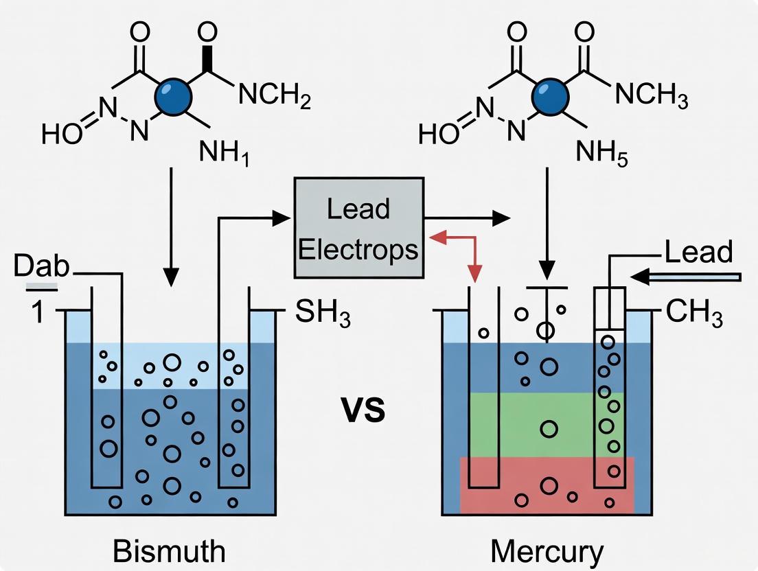
The accurate detection of lead (Pb²⁺) ions in environmental and biological samples is a critical analytical challenge due to the severe health risks posed by this toxic heavy metal. For decades, mercury-based electrodes have been considered the gold standard in anodic stripping voltammetry (ASV) for trace metal analysis due to their excellent electrochemical properties. However, with growing concerns about mercury's toxicity, bismuth-based electrodes have emerged as a promising environmentally friendly alternative. The fundamental interaction between these electrode materials and lead ions—specifically their alloying mechanisms—plays a pivotal role in determining the sensitivity, selectivity, and overall performance of electrochemical sensors.
Both bismuth and mercury function effectively in stripping voltammetry because they form alloys with lead and other heavy metals during the analysis process. This alloy formation concentrates the target metals into the electrode surface, enabling the highly sensitive detection required for environmental monitoring and biomedical applications. As research continues to refine these electrode materials, understanding their distinct alloying behaviors with lead provides crucial insights for sensor design and operation. This article systematically compares the alloying mechanisms of bismuth and mercury with lead ions, supported by experimental data and performance metrics from current research.
Fundamental Alloying Mechanisms
The interaction between electrode materials and lead ions during anodic stripping voltammetry follows a well-defined electrochemical pathway involving distinct deposition and stripping stages. The fundamental process can be visualized as follows:
Bismuth-Lead Alloying Mechanism
Bismuth film electrodes operate through the formation of binary alloys with lead and other metal ions during the deposition step. Research has confirmed that bismuth forms well-defined intermetallic compounds with lead, cadmium, thallium, and indium, where the metals are integrated into the bismuth crystal lattice structure. The resulting alloys exhibit distinct electrochemical signatures during the stripping step, producing sharp, well-defined peaks that enable highly sensitive detection of trace metals. This alloying behavior is functionally analogous to that of mercury, but with the significant advantage of bismuth's lower toxicity [20].
The bismuth-lead alloy formation is particularly favorable because it produces undistorted stripping peaks with excellent resolution between different metal species. This characteristic allows for convenient multi-element measurements down to the low μg/L level. One key limitation, however, is that copper does not form a binary alloy with bismuth, which can affect measurements in samples containing both copper and lead. This challenge can be circumvented by adding gallium to the solution, similar to approaches used with mercury film electrodes [20].
Mercury-Lead Alloying Mechanism
Mercury electrodes form amalgams with lead ions during the deposition phase of anodic stripping voltammetry. In this process, reduced lead atoms dissolve into the mercury matrix to form a mercury-lead amalgam. The homogeneous distribution of lead within the mercury film allows for highly efficient preconcentration of the target analyte. During the stripping phase, the lead is re-oxidized from the amalgam, producing characteristic current peaks that are proportional to the lead concentration in the original sample [21].
The mercury-lead amalgamation provides exceptional reproducibility and sensitivity, which has established mercury as the traditional reference material for stripping voltammetry. However, the toxicity of mercury presents significant handling, disposal, and environmental concerns that have motivated the search for alternative electrode materials. Additionally, mercury electrodes have a relatively limited anodic potential window, which can restrict their application for certain analytical challenges [1] [20].
Performance Comparison and Experimental Data
Quantitative Performance Metrics
Extensive research has compared the analytical performance of bismuth and mercury electrodes for lead detection. The following table summarizes key performance parameters derived from experimental studies:
Table 1: Performance comparison of bismuth vs. mercury electrodes for lead detection
| Performance Parameter | Bismuth Electrodes | Mercury Electrodes |
|---|---|---|
| Detection Limit | 0.001 μM (with Bi₂O₃/IL/rGO nanocomposite) [18] | Comparable sub-μg/L levels achievable [21] |
| Linear Range | 0.1-10 μg/mL (with mercury films) [1] | Wide linear response range demonstrated [21] |
| Peak Resolution | Well-defined, sharp peaks; excellent for neighboring signals [20] | Excellent peak resolution and shape [20] |
| Multi-element Capability | Effective for Cd(II), Pb(II), In(III); Cu(II) problematic [1] [20] | Comprehensive multi-element capability [20] |
| Toxicity | Low toxicity; "green" alternative [22] [20] | High toxicity; significant handling concerns [1] [20] |
| Electrode Stability | Good stability with proper modification; long-term degradation possible [18] | Consistent performance but prone to oxidation [23] |
| Reproducibility | High reproducibility with optimized films [20] | Excellent reproducibility [21] |
Advanced Bismuth-Based Nanocomposites
Recent developments in nanomaterial science have significantly enhanced the performance of bismuth-based electrodes. Researchers have created innovative hybrid nanocomposites such as bismuth oxide/ionic liquid/reduced graphene oxide (Bi₂O₃/IL/rGO) to overcome the limitations of pure bismuth films. These advanced materials demonstrate exceptional performance for lead detection, with a low detection limit of 0.001 μM and quantification limit of 0.003 μM [18].
The synergistic combination of materials in this nanocomposite addresses key limitations of bismuth electrodes: the ionic liquid provides high ionic conductivity and stabilization, reduced graphene oxide offers a large surface area and excellent electron transfer capability, and bismuth oxide enables effective alloy formation with lead ions. When tested on real environmental samples, sensors based on this nanocomposite showed acceptable recovery rates ranging from 95% to 102%, confirming their practical utility for environmental monitoring applications [18].
Experimental Protocols and Methodologies
Electrode Preparation and Modification
Bismuth Film Electrode Preparation
The preparation of bismuth film electrodes typically follows an ex situ electrodeposition approach. In a standard protocol, bismuth films are formed by applying a negative potential (typically -1.2 V to -1.4 V vs. Ag/AgCl) for 60-300 seconds in a solution containing 100-400 mg/L bismuth ions in an acetate buffer (pH 4.0-4.5) with 0.5 M sodium sulfate as supporting electrolyte. The deposition is performed under stirred conditions to ensure homogeneous film formation. The resulting bismuth film exhibits uniform coverage and provides an effective platform for subsequent lead detection [1] [20].
For paper-based bismuth electrodes, researchers have developed a specialized fabrication process involving wax-printed chromatography paper modified with carbon ink. The bismuth film is then electrodeposited onto this conductive paper substrate, creating a low-cost, disposable sensor platform ideal for field applications. This approach significantly reduces the amount of bismuth required while maintaining excellent analytical performance [1].
Mercury Film Electrode Preparation
Mercury film electrodes are typically prepared by electrodepositing mercury onto various substrates (most commonly glassy carbon) from a solution containing mercury ions. A standard protocol involves applying a deposition potential of -1.0 V to -1.2 V (vs. Ag/AgCl) for 300-600 seconds in a solution of 100-500 mg/L mercury acetate in 0.1 M HCl or 0.1 M nitrate solution. The thickness of the mercury film can be controlled by varying the deposition time and mercury ion concentration in the solution. The resulting electrode provides a renewable surface for repeated measurements, though the toxicity of mercury requires careful handling and disposal procedures [21].
Nanocomposite-Modified Electrodes
Advanced sensor architectures incorporate bismuth into nanocomposite structures to enhance performance. The synthesis of Bi₂O₃/IL/rGO nanocomposite involves dispersing 100 mg of graphene oxide (GO) in 100 mL deionized water through ultrasonication for 30 minutes. Simultaneously, a bismuth solution is prepared by dissolving bismuth nitrate pentahydrate (Bi(NO₃)₃·5H₂O) in deionized water. These solutions are combined, followed by the addition of ionic liquid (1-butyl-3-methylimidazolium hexafluorophosphate, BMIM-PF6). The mixture undergoes vigorous stirring and is then transferred to a Teflon-lined autoclave for hydrothermal treatment at 120-180°C for 6-12 hours. The resulting precipitate is collected, washed, and dried to obtain the final Bi₂O₃/IL/rGO nanocomposite [18].
For electrode modification, the nanocomposite is dispersed in a suitable solvent (often with the addition of Nafion as a binder), and a precise volume (typically 2-10 μL) is drop-cast onto the electrode surface (e.g., glassy carbon electrode). The modified electrode is then dried at room temperature or under mild heating to form a stable sensing film [18].
Analytical Measurement Procedures
Anodic Stripping Voltammetry Protocol
Anodic stripping voltammetry for lead detection follows a standardized sequence, as visualized below:
For bismuth film electrodes, the analytical procedure typically employs square wave anodic stripping voltammetry with the following parameters: deposition potential of -1.2 V, deposition time of 120-300 seconds (depending on lead concentration), equilibration time of 15 seconds, square wave amplitude of 25 mV, frequency of 15 Hz, and step potential of 5 mV. The stripping scan typically ranges from -1.2 V to -0.2 V, with lead producing a well-defined peak at approximately -0.5 V (vs. Ag/AgCl) [20].
For mercury film electrodes, a similar approach is used, though the deposition potential may be slightly less negative (-1.0 V to -1.1 V) to prevent hydrogen evolution. The stripping scan typically covers a range from -1.0 V to -0.1 V, with lead appearing at a similar potential of approximately -0.5 V. The choice of stripping technique (square wave vs. differential pulse) can be optimized based on the specific sample matrix and interference considerations [21].
The Scientist's Toolkit: Essential Research Reagents and Materials
Table 2: Essential research reagents and materials for bismuth and mercury electrode experiments
| Reagent/Material | Function in Experiment | Typical Concentration/Form |
|---|---|---|
| Bismuth nitrate pentahydrate | Source of Bi³⁺ ions for film formation | 100-1000 mg/L in deposition solution [1] |
| Mercury(II) acetate | Source of Hg²⁺ ions for film formation | 100-500 mg/L in 0.1 M HCl [21] |
| Acetate buffer | pH control and supporting electrolyte | 0.1 M, pH 4.0-4.5 [20] |
| Sodium sulfate | Supporting electrolyte for conductivity | 0.5 M in deposition solution [1] |
| 1-Butyl-3-methylimidazolium hexafluorophosphate | Ionic liquid for nanocomposite stabilization | 0.5-2% in nanocomposite synthesis [18] |
| Graphene oxide | Nanomaterial backbone for composites | 100 mg/100 mL in synthesis [18] |
| Nafion solution | Binder for electrode modification | 0.1% dilution for drop-casting [18] |
| Lead nitrate | Standard solution for calibration | 1000 mg/L stock, diluted to working concentrations [18] |
The alloying mechanisms of bismuth and mercury with lead ions share fundamental similarities that enable highly sensitive detection in anodic stripping voltammetry. Both materials form defined metallic phases with lead—bismuth as binary alloys and mercury as amalgams—that concentrate the analyte during deposition and release it during the stripping step to produce quantifiable current signals. Experimental data demonstrates that properly optimized bismuth-based electrodes can achieve performance comparable to traditional mercury electrodes, with detection limits reaching 0.001 μM for advanced nanocomposites.
The choice between bismuth and mercury electrodes involves balancing multiple factors including toxicity, sensitivity, reproducibility, and application requirements. While mercury electrodes remain a robust reference standard with proven performance across diverse matrices, bismuth electrodes offer a compelling "green" alternative with significantly reduced toxicity concerns. Ongoing research in nanomaterial engineering, particularly the development of bismuth-based nanocomposites, continues to enhance the capabilities of bismuth electrodes, positioning them as the leading sustainable technology for future electrochemical monitoring of lead and other toxic heavy metals.
Modern Sensor Designs and Methodologies for Real-World Application
The bismuth film electrode (BiFE) has emerged as a leading environmentally friendly alternative to traditional mercury-based electrodes for the electrochemical detection of trace heavy metals. [24] [2] Since its introduction in 2000, BiFE has attracted significant attention in stripping analysis due to its low toxicity, well-defined stripping signals, and insensitivity to dissolved oxygen. [24] A critical consideration in implementing this technology is the film preparation method, primarily classified as in-situ (where the bismuth film is plated simultaneously with the target metals during analysis) or ex-situ (where the bismuth film is pre-plated in a separate step before transfer to the measurement solution). [24] This guide provides an objective comparison of these two configurations, framing the discussion within the broader performance context of bismuth versus mercury electrodes for lead detection research.
Fundamental Principles and Comparison to Mercury Electrodes
Bismuth film electrodes function by forming "fused alloys" with heavy metals such as lead (Pb), cadmium (Cd), and zinc (Zn) during the preconcentration step of anodic stripping voltammetry (ASV). [25] [2] This process is analogous to the amalgamation process at mercury electrodes, but utilizes a material with vastly lower toxicity. [26] [2] The attractive properties of BiFEs include high sensitivity, excellent peak resolution, a wide operational potential window, and the fact that they do not require deaeration of the solution, which is a significant practical advantage for on-site monitoring. [24] [2]
The motivation for replacing mercury electrodes is strongly supported by health and safety evidence. Mercury is a potent neurotoxin, and exposure—particularly to inhaled mercury vapor—can cause tremors, emotional changes, insomnia, neuromuscular changes, and cognitive deficits. [27] [28] Reports of occupational mercury exposure in recycling facilities highlight the serious risks, with workers showing elevated urine mercury levels and symptoms consistent with mercury toxicity. [27]
The following diagram illustrates the general workflow for preparing and using bismuth film electrodes, highlighting the key decision point between in-situ and ex-situ methods.
Performance Comparison: In-Situ vs. Ex-Situ Bismuth Films
The choice between in-situ and ex-situ preparation significantly impacts analytical performance, practicality, and suitability for specific applications. The table below summarizes the core characteristics of each method.
Table 1: Direct comparison of in-situ and ex-situ bismuth film preparation methods
| Feature | In-Situ Bismuth Film | Ex-Situ Bismuth Film |
|---|---|---|
| Preparation Principle | Bi(III) ions added to sample; simultaneous co-deposition of Bi film and target metals during preconcentration. [14] [26] | Bi film pre-plated onto substrate electrode in a separate solution before transfer to measurement cell. [24] |
| Procedure Simplicity | Simple; fewer steps, no transfer of filmed electrode required. [14] | More complex; requires a separate plating step and careful electrode transfer. [24] |
| Film Stability/Adhesion | Good; "better adhesion of the Bi film to the... surface" due to simultaneous deposition. [26] | Can be problematic; severe film stability problems were observed on microelectrodes, requiring careful optimization. [24] |
| Analytical Sensitivity | High; "co-deposition of Bi(III) and Cd2+ can enhance the enrichment of Cd2+ by forming Bi-Cd alloy, thus improving the detection sensitivity." [14] | High and stable; with optimization, offers "excellent long-term film functional stability" and "attractive stripping analytical performance." [24] |
| Operational Flexibility | Lower; the same film is used for a single measurement. Requires Bi(III) in measurement solution. | Higher; the same pre-plated film can be used for multiple measurement events. [24] |
| Preferred Application Context | Routine analysis in well-defined solutions where reagent addition is acceptable; food, environmental water. [14] [2] | Micro-analysis, small volumes, in-vivo measurements, adsorptive stripping where Bi(III) interferes, flow detectors. [24] |
Experimental Performance Data
To support the selection process, the following table compiles key analytical figures of merit reported in the literature for the detection of lead (Pb) and other metals using both configurations.
Table 2: Experimental performance data for heavy metal detection using different bismuth film configurations
| Electrode Configuration | Target Analyte | Limit of Detection (LOD) | Linear Range | Reproducibility (RSD) | Key Experimental Conditions | Citation |
|---|---|---|---|---|---|---|
| In-situ Bi/SPCE | Cd(II) | 3.55 µg/L | 5–100 µg/L | N/R | Acetate buffer, 180s deposition, SWASV [14] | |
| In-situ Bi/Pencil Graphite | Pb(II) & Cd(II) | 11.5 µg/L (Pb), 11.0 µg/L (Cd) | N/R | N/R | Acetate buffer (pH 4.5), 250s deposition, DPASV [26] | |
| Ex-situ Bi/Carbon Fibre | Pb(II) | ~0.1 µg/L (as part of Cd/Pb mix) | Demonstrated for 10 µg/L | ~2.4% (for Cd/Pb) | Acetate buffer, 120s preconcentration, ASV [24] | |
| Nafion-coated In-situ Bi/GC | Pb(II) | 0.17 µg/L | N/R | 2.4% (at 15 µg/L, n=15) | Acetate buffer, 180s preconcentration, DPASV [2] | |
| Ex-situ Bi/Carbon Fibre (AdCSV) | Ni(II) | 90 ng/L | Highly linear | 2.9% (at 1 µg/L, n=10) | Ammonia buffer, 120s preconcentration, AdCSV [24] |
N/R: Not explicitly Reported in the cited source.
Detailed Experimental Protocols
Protocol for In-Situ Bismuth Film Electrode Preparation
This protocol is adapted from studies using screen-printed carbon electrodes (SPCEs) for cadmium and lead detection. [14] [26]
- Electrode Pre-treatment (Pre-anodization - Optional but recommended): To enhance electron transfer rate, pre-anodize the screen-printed carbon electrode by cyclic voltammetry. Scan the SPCE for 5 cycles in 0.1 mol/L PBS (pH = 9) between 0.5 V and 1.7 V at a scan rate of 0.1 V/s. Rinse thoroughly with ultrapure water and dry at room temperature. [14]
- Solution Preparation: Prepare a measurement solution containing your sample or standard, a supporting electrolyte (e.g., 0.1 M acetate buffer, pH 4.5), and a Bi(III) ion source (e.g., 150-250 mg/L Bi(III) from Bi(NO~3~)~3~ stock solution). Sodium bromide (20 µM) may be added as a plating agent. [14] [26]
- Analysis via SWASV/DPASV:
- Deposition/Preconcentration: Apply a deposition potential of -1.4 V to -1.5 V vs. Ag/AgCl for 180-250 s under stirring. During this step, Bi(III) and target metal ions (e.g., Cd²⁺, Pb²⁺) are co-deposited onto the electrode surface, forming the bismuth film and alloys simultaneously.
- Equilibration: Stop stirring and allow the solution to equilibrate for ~15 s.
- Stripping: Record the anodic stripping voltammogram using Square-Wave (SWASV) or Differential Pulse (DPASV) modes by scanning from the deposition potential to a more positive potential (e.g., -0.2 V). The oxidation (stripping) of the target metals produces characteristic current peaks.
Protocol for Ex-Situ Bismuth Film Electrode Preparation
This protocol is based on the optimized procedure for preparing ex-situ bismuth film microelectrodes (BiFMEs) on carbon fiber substrates. [24]
- Substrate Electrode Preparation: Polish and clean the substrate electrode (e.g., carbon fiber, glassy carbon) according to standard procedures.
- Plating Solution Preparation: Prepare a separate plating solution containing 0.1 M acetate buffer (pH 4.5), 250 mg/L Bi(III), and 0.1 M NaBr, which acts as a plating agent crucial for forming a stable film. [24]
- Film Electrodeposition: Immerse the substrate electrode into the plating solution. Apply an optimized deposition potential of -1.0 V vs. Ag/AgCl for 300 s under stirring to electroplate the bismuth film onto the substrate.
- Electrode Transfer: Carefully remove the prepared bismuth film electrode (BiFE) from the plating solution, rinse gently with ultrapure water, and transfer it to the measurement cell (sample solution).
- Analysis via Stripping Voltammetry: The measurement solution contains the target analytes and supporting electrolyte but no Bi(III) ions. Perform the stripping voltammetry procedure (deposition and stripping) as described for the in-situ method. The same ex-situ plated film can be used for multiple measurements. [24]
The Scientist's Toolkit: Essential Research Reagents and Materials
Table 3: Key reagents and materials for bismuth film electrode experiments
| Item | Function/Description | Example in Context |
|---|---|---|
| Bismuth Salt | Source of Bi(III) ions for film formation. | Bismuth nitrate pentahydrate (Bi(NO₃)₃·5H₂O) is commonly used to prepare stock solutions. [26] |
| Supporting Electrolyte | Provides ionic conductivity and controls pH. | Acetate buffer (0.1 M, pH 4.5) is widely used for the detection of Cd and Pb. [14] [26] |
| Plating Agent | Enhances the quality and stability of the ex-situ plated bismuth film. | Sodium Bromide (NaBr, 0.1 M) in the plating solution was critical for achieving a stable ex-situ film. [24] |
| Substrate Electrodes | The base conductor on which the bismuth film is deposited. | Carbon-based materials are most common: Glassy Carbon (GC), Screen-Printed Carbon Electrodes (SPCE), Carbon Fiber, Pencil Graphite. [24] [14] [26] |
| Nafion Polymer | A cation-exchange polymer coating used to modify the electrode surface to improve selectivity and antifouling properties. | A Nafion-coated bismuth film electrode (NCBFE) was used for the determination of heavy metals in vegetable samples. [2] |
Both in-situ and ex-situ bismuth film electrode configurations offer compelling, high-performance alternatives to mercury electrodes, aligning with modern demands for environmentally friendly analytical chemistry. The in-situ method is generally simpler and benefits from fresh film formation for each analysis, often yielding excellent sensitivity, making it ideal for most routine laboratory analyses. In contrast, the ex-situ method, while requiring more meticulous optimization, provides superior mechanical stability and operational flexibility, which is critical for applications involving micro-volumes, flow systems, or multiple analyses with a single film. The choice is not about which is universally better, but which is more appropriate for the specific analytical challenge and experimental constraints.
The detection of toxic heavy metals, such as lead, is a critical priority in environmental monitoring, food safety, and healthcare. For decades, mercury-based electrodes were the gold standard for this application due to their excellent electrochemical properties, including a wide cathodic window, high reproducibility, and the ability to form amalgams with metals [6]. However, the high toxicity of mercury has driven the scientific community to seek safer, environmentally friendly alternatives [6]. Bismuth has emerged as the most viable successor, offering a comparable ability to form alloys with heavy metals, a wide potential window, low background current, and very low toxicity [11] [6]. This review objectively compares the performance of advanced bismuth-composite electrodes against traditional mercury electrodes and delineates the experimental protocols used for their evaluation in lead detection research.
Performance Comparison: Bismuth-Composite vs. Mercury Electrodes
The development of bismuth-based electrodes has evolved from pure bismuth films to sophisticated composites integrating nanomaterials and conductive polymers. These advanced materials aim to overcome the historical limitations of bismuth, such as susceptibility to hydrolysis in alkaline conditions and fouling in complex media [11]. The table below summarizes key performance metrics for different electrode types in the detection of lead and other heavy metals.
Table 1: Performance Comparison of Mercury, Plain Bismuth, and Advanced Bismuth-Composite Electrodes for Heavy Metal Detection
| Electrode Type | Target Analytes | Linear Range (µg/mL) | Limit of Detection (LOD, µg/mL) | Key Advantages | Key Limitations |
|---|---|---|---|---|---|
| Mercury Film [6] | Cd(II), Pb(II), In(III), Cu(II) | 0.1 - 10 | Pb(II): 0.1 | High sensitivity, well-established method | High toxicity, environmental and health hazards |
| Plain Bismuth Film [6] | Cd(II), Pb(II), In(III) | Not Specified | Pb(II): Comparable to Mercury | Low toxicity, "environmentally friendly" | Cannot detect Cu(II); prone to fouling |
| Antifouling Bismuth Composite (BSA/g-C₃N₄/Bi₂WO₆/GA) [11] | Multiple Heavy Metals | Not Specified | Not Specified | Retains 90% signal after 1 month in biofluids/wastewater; Robust in complex matrices | More complex fabrication process |
| Paper-based Bi Film [6] | Cd(II), Pb(II) | Not Specified | Not Specified | Low-cost, disposable, portable | Potentially lower reproducibility |
Experimental data confirms that mercury films offer a slightly superior analytical performance, with low limits of detection for a wider range of metals, including copper [6]. However, plain bismuth films provide a more sustainable alternative with comparable sensitivity for lead and cadmium. The most significant recent advancement is in antifouling bismuth composites. One study demonstrated that a composite of bismuth tungstate (Bi₂WO₆) integrated into a 3D porous matrix of cross-linked bovine serum albumin (BSA) and 2D graphitic carbon nitride (g-C₃N₄) retained over 90% of its electrochemical signal after one month of storage in challenging complex matrices like untreated human plasma, serum, and wastewater [11]. This addresses a major commercialization hurdle for bismuth-based sensors: performance degradation in real-world samples.
Experimental Protocols for Electrode Fabrication and Testing
Fabrication of Antifouling Bismuth-Composite Electrodes
The protocol for creating the robust BSA/g-C₃N₄/Bi₂WO₆/GA coating is as follows [11]:
- Solution Preparation: A pre-polymerization solution is prepared using BSA and g-C₃N₄ as functional monomers, glutaraldehyde (GA) as a cross-linker, and flower-like bismuth tungstate (Bi₂WO₆) as a heavy metal co-deposition anchor.
- Mixing: The solution is homogenized through mixing and ultrasonic treatment to ensure uniform dispersion.
- Coating Formation: The solution is immediately drop-cast onto the surface of a gold or other suitable electrode.
- Polymerization: The coating is allowed to polymerize and form a stable, cross-linked, 3D porous sponge-like matrix on the electrode surface. Characterization via Scanning Electron Microscopy (SEM) confirms the porous structure, while X-ray Photoelectron Spectroscopy (XPS) verifies the successful polymerization.
Fabrication of Paper-Based Bismuth Film Electrodes
A low-cost and disposable alternative uses paper-based substrates [6]:
- Substrate Preparation: Paper-based carbon working electrodes are fabricated, for instance, by depositing carbon ink on wax-printed paper to define the electrode area.
- Film Deposition (Ex Situ): The paper-based working electrode is placed in an electrochemical cell. A bismuth film is electrodeposited onto the carbon surface from a separate solution containing a bismuth salt (e.g., 10⁻³ M Bismuth in acetate buffer with 0.5 M Na₂SO₄ as a supporting electrolyte) by applying a negative potential.
Analytical Measurement via Anodic Stripping Voltammetry
The core experimental protocol for detecting lead and other heavy metals is anodic stripping voltammetry, which follows these steps for both mercury and bismuth-based electrodes [6]:
- Preconcentration/Electrodeposition: The modified electrode is placed in a stirred sample solution containing the target metal ions. A negative potential is applied, reducing the metal ions (e.g., Pb²⁺) to their zero-valent state (Pb⁰) and forming an alloy with the bismuth (or amalgam with mercury) on the electrode surface.
- Equilibration: The stirring is stopped, and the solution is allowed to become quiescent.
- Stripping: An anodic (positive-going) potential sweep is applied. This re-oxidizes the deposited metals, causing them to strip back into the solution. Each metal strips at a distinct characteristic potential.
- Quantification: The resulting current peak is measured. The peak height (current) is proportional to the concentration of the metal in the original sample.
The following diagram illustrates this core experimental workflow:
Diagram 1: Anodic Stripping Voltammetry Workflow
The Scientist's Toolkit: Essential Research Reagents and Materials
Successful research and application in this field rely on a specific set of materials. The table below lists key reagents, their functions, and examples from the literature.
Table 2: Essential Research Reagents for Bismuth-Composite Electrode Development
| Reagent/Material | Function in Experiment | Specific Examples |
|---|---|---|
| Bismuth Precursors | Source of bismuth for forming the electroactive film or composite. | Bismuth nitrate pentahydrate (Bi(NO₃)₃·5H₂O) [29], Bismuth standard solutions for ICP [6]. |
| Conductive Polymers | Enhance electron transfer, provide a matrix for biocomponent entrapment, and improve sensitivity. | Polypyrrole (PPy), Polyaniline (PANI), Poly(3,4-ethylenedioxythiophene) (PEDOT) [30] [31]. |
| 2D Nanomaterials | Increase surface area, enhance electrical conductivity, and provide anchoring sites for metal ions. | Graphitic Carbon Nitride (g-C₃N₄) [11], Reduced Graphene Oxide (RGO) [29]. |
| Cross-linking Agents | Form stable, 3D porous polymer networks that encapsulate functional materials and provide antifouling properties. | Glutaraldehyde (GA) [11]. |
| Proteins & Ligands | Act as ion-selective chelators or form biocompatible, antifouling matrices. | Bovine Serum Albumin (BSA) [11]. |
| Supporting Electrolytes | Provide ionic strength and conductivity in the solution, enabling electrochemical reactions. | Acetate buffer, Sodium Sulfate (Na₂SO₄) [6]. |
Signaling Pathways in Bismuth-Composite Induced Toxicity
While bismuth is low-toxicity, understanding the biological interactions of its nano-composites is crucial for safe application, especially in biomedical contexts. Studies on bismuth oxide/reduced graphene oxide (Bi₂O₃/RGO) nanocomposites in mammalian cell lines have revealed a toxicity mechanism primarily driven by oxidative stress.
The following diagram illustrates the apoptotic signaling pathway induced in cells exposed to Bi₂O₃/RGO nanocomposites:
Diagram 2: Toxicity Pathway of Bi₂O₃/RGO Nanocomposites
This mechanistic insight is vital for designing safe bismuth composites. The observed cytotoxic effects are dose- and time-dependent, with one study showing normal rat kidney cells (NRK52E) were marginally more vulnerable than human liver cancer cells (HepG2) [29].
The transition from mercury to bismuth-based electrodes represents a significant advancement in sustainable electroanalysis. While plain bismuth films offer a non-toxic alternative with performance comparable to mercury for key analytes like lead, the future lies in advanced bismuth composites. The integration of bismuth with conductive polymers and nanostructured materials like g-C₃N₄ and RGO directly addresses the critical challenges of sensitivity, selectivity, and robustness in complex real-world samples. Experimental data confirms that these novel composites, particularly those engineered with antifouling properties, can maintain performance in environments where traditional electrodes fail, thereby closing the performance gap with mercury and opening new avenues for commercial application in healthcare, environmental monitoring, and food safety.
The environmental monitoring of toxic heavy metals, such as lead and cadmium, is of critical importance due to their toxicity, non-biodegradability, and ability to accumulate in living tissues [7]. For decades, mercury electrodes were the gold standard in stripping voltammetry for trace metal detection due to their excellent electrochemical properties, including a wide cathodic window and reproducible surface [6]. However, the high toxicity of mercury has triggered intensive research into safer, more environmentally friendly alternatives [7] [6].
Bismuth has emerged as the most promising substitute, offering comparable analytical performance with very low toxicity [32] [6]. Concurrently, the field has witnessed a trend toward miniaturization and solid-state platforms, including microelectrode arrays and paper-based sensors, which provide enhanced portability, reduced sample/reagent consumption, and suitability for decentralized analysis [7] [33]. This guide objectively compares the analytical performance of these emerging bismuth-based platforms against traditional mercury-based systems, providing experimental data and methodologies to inform researcher selection for specific applications.
Performance Comparison: Bismuth vs. Mercury Electrodes
The following tables summarize key performance metrics for bismuth and mercury-based sensors in the detection of heavy metals, particularly lead (Pb) and cadmium (Cd).
Table 1: Overall Performance Comparison of Bismuth vs. Mercury Electrodes
| Parameter | Bismuth-Based Electrodes | Mercury-Based Electrodes |
|---|---|---|
| Toxicity & Environmental Impact | Very low toxicity; more environmentally friendly [6]. | Highly toxic; requires special handling and disposal [7] [6]. |
| Primary Form | Solid bismuth microelectrode arrays, bismuth film electrodes (ex situ or in situ) [7] [6]. | Mercury films electrodeposited on carbon substrates [6]. |
| Sensitivity (Example for Pb/Cd) | LOD for Pb(II): 8.9×10⁻¹⁰ mol L⁻¹; for Cd(II): 2.3×10⁻⁹ mol L⁻¹ (Solid Bi μEA) [7]. | LOD for Pb(II): 0.1 µg/mL (~4.8×10⁻¹⁰ mol L⁻¹); for Cd(II): 0.4 µg/mL (~3.6×10⁻⁹ mol L⁻¹) (Hg-film paper electrode) [6]. |
| Linear Range (Example for Pb/Cd) | Cd(II): 5×10⁻⁹ to 2×10⁻⁷ mol L⁻¹; Pb(II): 2×10⁻⁹ to 2×10⁻⁷ mol L⁻¹ [7]. | 0.1 to 10 µg/mL for Cd(II), Pb(II), In(III), and Cu(II) [6]. |
| Metal Detection Scope | Cd(II), Pb(II), In(III); Cu(II) could not be determined with bismuth films on paper [6]. | Cd(II), Pb(II), In(III), Cu(II) (wider scope) [6]. |
Table 2: Comparison of Miniaturized Bismuth Platforms
| Platform | Key Features | Analytical Performance | Advantages |
|---|---|---|---|
| Solid Bismuth Microelectrode Array [7] | - 43 single capillaries (d~10 µm) filled with metallic Bi- Reusable design- No need for Bi(III) addition | - LOD (Pb): 8.9×10⁻¹⁰ mol L⁻¹- LOD (Cd): 2.3×10⁻⁹ mol L⁻¹- RSD: ~4.1% (for Sunset Yellow dye) [34] [35] | - Eco-friendly- Amplified currents- Microelectrode properties (spherical diffusion)- Long-term use [7] [35] |
| Paper-based Carbon Electrode with Bi Film [6] | - Ex-situ electrodeposited Bi film- Low-cost, disposable substrate- Foldable and portable | - LODs in the µg/mL range (less sensitive than μEA)- Linear range: 0.1-10 µg/mL | - Extremely low cost- Easy disposal- Suitable for resource-limited settings [33] [6] |
| Lithographically Fabricated Bi μEA [32] | - Bi microdisk arrays made via sputtering/microlithography- Disposable sensor | - Well-defined signals for Cd(II) and Pb(II) in unstirred solutions | - No conductive substrate needed- Uniform, reproducible surface- Enhanced analytics in unstirred solutions [32] |
Experimental Protocols for Key Platforms
Solid Bismuth Microelectrode Array for Cd and Pb Detection
This procedure utilizes a reusable array of forty-three bismuth microelectrodes for the simultaneous determination of cadmium and lead via Anodic Stripping Voltammetry (ASV) [7].
- 1. Apparatus and Reagents: Autolab PGSTAT 10 potentiostat or equivalent. Acetate buffer (0.05 M, pH 4.6) as supporting electrolyte. Standard solutions of Cd(II) and Pb(II). The solid bismuth microelectrode array, Ag/AgCl reference electrode, and platinum auxiliary electrode [7].
- 2. Electrode Activation: Begin with an activation step by applying a potential of -2.75 V for 2 seconds to reduce surface oxides and ensure a fresh, metallic bismuth surface [7] [35].
- 3. Preconcentration/Deposition: Immerse the electrode in a stirred sample solution containing the target analytes. Apply a deposition potential of -1.4 V (vs. Ag/AgCl) for 60 seconds. During this step, Cd²⁺ and Pb²⁺ ions are reduced and amalgamated into the bismuth surface [7].
- 4. Stripping Scan: After a 5-second equilibration period, perform an anodic scan from -1.4 V to -0.2 V using Square-Wave Voltammetry (SWV). The amalgamated metals are re-oxidized, producing characteristic current peaks at -0.8 V for Cd and -0.55 V for Pb (vs. Ag/AgCl) [7].
- 5. Calibration and Quantification: Record the stripping peaks for a series of standard solutions to create a calibration curve. The peak current is proportional to the metal ion concentration in the sample [7].
Diagram 1: ASV Workflow for Solid Bismuth Microelectrode Array.
Paper-Based Electrode with Mercury vs. Bismuth Films
This protocol compares the ex-situ formation of mercury and bismuth films on low-cost paper-based carbon electrodes [6].
- 1. Fabrication of Paper-Based Working Electrode: Create hydrophobic barriers on chromatography paper using a wax printer and heat treatment. Prepare carbon ink by mixing carbon paste with N,N-dimethylformamide (DMF). Deposit the ink onto the defined paper zone using a spray adhesive and a shadow mask, then cure [6].
- 2. Ex-Situ Film Deposition: For Mercury Film: Place the paper electrode in a cell containing 10⁻³ M Hg(II) acetate in 0.1 M HCl. Apply a potential of -0.9 V for 300 seconds to electrodeposit the mercury film. For Bismuth Film: Place the electrode in a cell containing 10⁻³ M Bi(III) in acetate buffer (pH 4.0). Apply a potential of -1.0 V for 120 seconds to electrodeposit the bismuth film [6].
- 3. Analytical Measurement via ASV: Transfer the modified paper electrode to a new cell containing the sample in acetate buffer (pH 4.0) with 0.5 M Na₂SO₄. Apply a deposition potential (e.g., -1.4 V) for a set time with stirring. Follow with an anodic potential scan. The paper electrode is disposable after a single measurement [6].
The Scientist's Toolkit: Key Research Reagent Solutions
Table 3: Essential Reagents and Materials for Sensor Fabrication and Analysis
| Reagent/Material | Function/Application | Example Use Case |
|---|---|---|
| Metallic Bismuth (Solid) | Core electrode material for solid microelectrode arrays [7]. | Fabrication of the 43-capillary solid bismuth microelectrode array [7]. |
| Bismuth Salts (e.g., Bi(III) for ICP) | Precursor for electroplating bismuth films onto substrate electrodes [6]. | Preparation of 10⁻³ M Bi(III) solution in acetate buffer for ex-situ bismuth film deposition on paper electrodes [6]. |
| Mercury(II) Acetate | Precursor for electroplating mercury films (toxic, requires careful handling) [6]. | Preparation of 10⁻³ M Hg solution in 0.1 M HCl for ex-situ mercury film deposition for performance comparison studies [6]. |
| Acetate Buffer (pH ~4.6) | Common supporting electrolyte for ASV of heavy metals like Cd and Pb [7] [6]. | Optimal electrolyte (0.05 M) for the simultaneous determination of Cd(II) and Pb(II) using the solid bismuth microelectrode array [7]. |
| Sodium Sulfate (Na₂SO₄) | Background electrolyte to increase ionic strength and conductivity [6]. | Used at 0.5 M concentration in acetate buffer for analysis with paper-based film electrodes [6]. |
| Carbon Paste/Ink | Conductive substrate for fabricating low-cost electrodes, including paper-based sensors [6]. | Formulation of the conductive surface on paper-based working electrodes prior to bismuth or mercury film modification [6]. |
The experimental data confirms that bismuth-based sensors are a viable, high-performance, and eco-friendly alternative to mercury-based electrodes. Solid bismuth microelectrode arrays demonstrate superior sensitivity, achieving detection limits comparable to, and in some cases surpassing, those of mercury films (e.g., LOD for Pb(II) of 8.9×10⁻¹⁰ mol L⁻¹) [7]. Their key advantage lies in their microelectrode properties, which enable efficient mass transport via spherical diffusion, making them suitable for analysis in unstirred solutions and simplifying the measurement procedure [7]. Furthermore, their reusable design and elimination of the need to add Bi(III) ions to the sample make them more environmentally sustainable and suitable for long-term use [7] [35].
For applications where cost and disposability are paramount, paper-based bismuth film electrodes offer a compelling solution, though with a trade-off in sensitivity [6]. While mercury films on paper were found to be slightly more sensitive and capable of detecting a wider range of metals, including copper, bismuth films provide a significantly safer and more sustainable option for routine monitoring of Cd and Pb [6].
In conclusion, the choice between mercury and bismuth platforms depends on the specific analytical requirements. For ultra-trace analysis requiring the highest sensitivity and a broad metal detection scope, mercury may still be considered. However, for the vast majority of applications, particularly in environmental water monitoring, bismuth-based platforms—especially solid microelectrode arrays for lab-based analysis and paper-based sensors for field deployment—deliver a powerful, eco-friendly, and effective solution for heavy metal detection.
In electroanalysis, particularly for trace metal detection in complex matrices, the performance of a sensor is intrinsically linked to the properties of its surface. The electrode-solution interface is a critical battlefield where detection occurs, but also where interference accumulates. For sensors designed to detect heavy metals like lead in biofluids and environmental samples, non-specific adsorption of proteins, organic matter, and other matrix components can foul the electrode surface, leading to signal suppression, baseline drift, and irreproducible results. This matrix complexity poses a significant challenge for reliable analytical measurements.
The broader research on bismuth vs. mercury electrodes for lead detection provides a foundational perspective on interfacial design. While mercury film electrodes have long been the gold standard for anodic stripping voltammetry due to their high sensitivity and renewable surface, toxicity concerns have driven the search for safer alternatives. Bismuth film electrodes have emerged as a promising, environmentally friendly option with a stripping performance that compares favorably to their mercury counterparts [1] [20]. This comparison extends beyond mere toxicity to fundamental interfacial behavior. Both bismuth and mercury form alloys with heavy metals like lead, cadmium, and zinc during the preconcentration step, which is key to their excellent stripping performance [20]. Understanding how these electrode materials resist fouling in complex media is essential for developing robust sensors for real-world applications, bridging the gap between controlled laboratory experiments and analysis of intricate samples like blood, urine, or seawater.
Fundamental Mechanisms of Contaminant Adhesion
A deep understanding of the interaction between pollutants and an interface is crucial for the targeted development of effective antifouling strategies. The adhesion of contaminants is governed by a complex interplay of chemical, physical, and mechanical forces.
Chemical and Physical Interactions
The initial adhesion of contaminants is primarily mediated by chemical and physical forces. Chemical factors include covalent bonds, ionic bonds, and coordination bonds. For instance, proteins can bind to surfaces through thiol groups forming robust gold-thiol bonds, or amino groups can form Schiff bases with aldehyde-functionalized surfaces [36]. Mussels achieve remarkable underwater adhesion through the coordination of dopamine with metal ions [36]. Physical adsorption encompasses hydrogen bonding, van der Waals forces, and hydrophobic interactions. Long-chain fatty acids and lipids can displace interfacial water via hydrophobic interactions and then bind through van der Waals forces, forming tenacious films that facilitate further fouling [36].
Temporal Sequence of Fouling
The fouling process typically follows a distinct spatial and temporal sequence. In marine environments, the surface is first conditioned by an organic film of proteins and polysaccharides. This film creates a favorable microenvironment for bacterial colonization. These bacteria, in turn, secrete metabolic products and extracellular polymeric substances, altering the surface morphology and providing nutrients and adhesion sites for larger organisms like mussel larvae and barnacles [36]. A analogous process occurs in biofluids; for example, thrombus formation on medical devices is initiated by the adhesion of proteins like fibrinogen, followed by platelet attachment and activation [36]. This layered understanding of fouling mechanisms directly informs the design of antifouling coatings for electrochemical sensors, where preventing the initial organic conditioning film is paramount for maintaining signal fidelity.
Antifouling Strategies and Coating Mechanisms
Shielding adhesive forces and interrupting the contamination adhesion process are the core principles guiding antifouling coating research. Current strategies primarily leverage four key mechanisms to prevent the attachment or facilitate the removal of fouling agents.
Table 1: Core Antifouling Mechanisms and Their Principles
| Mechanism | Principle of Action | Common Coating Types |
|---|---|---|
| Surface Energy Tuning | Adjusts surface free energy to a range that minimizes interfacial adhesion, as described by the Baier curve [36]. | Silicone-based, Fluoropolymer |
| Superhydrophobic/Superhydrophilic Surfaces | Creates extreme wetting states: superhydrophobic surfaces trap air, preventing contact; superhydrophilic forms a hydrated layer that blocks adhesion [36]. | Fluoropolymer, Hydrogel |
| Surface Microstructuring | Introduces physical topographies that reduce the effective contact area for adhesives or create mechanically unstable points for settling organisms [36]. | Engineered polymers |
| Slippery Liquid-Infused Porous Surfaces (SLIPS) | Constructs a lubricant-infused, ultrasmooth surface that presents no stable anchor points for contaminants [36]. | Liquid-impregnated surfaces |
Performance Comparison: Biocidal vs. Biocide-Free Coatings
The choice between biocidal and biocide-free coatings represents a fundamental trade-off between traditional efficacy and modern environmental and safety considerations. This comparison is critical for applications ranging from ship hulls to specialized sensors.
Efficacy and Environmental Impact
Biocidal coatings, typically containing copper-based compounds like cuprous oxide or copper pyrithione, function by continuously leaching toxicants to deter the settlement and growth of fouling organisms [37] [38]. These coatings have demonstrated effective short-term fouling control and are the dominant market choice. However, a significant drawback is the release of these biocides into the surrounding environment, raising concerns about ecological harm and the potential for developing biocide-resistant species [39] [38]. In contrast, biocide-free coatings, such as silicone-based foul-release types, operate on a physical principle. They feature low surface energy and smooth, non-porous surfaces that minimize the adhesion strength of organisms, allowing them to be easily removed by hydrodynamic forces [37] [38]. These coatings offer a more environmentally benign profile, producing no toxic leachates.
Table 2: Comparison of Biocidal and Biocide-Free Antifouling Coatings
| Parameter | Biocidal Coatings | Biocide-Free (Foul-Release) Coatings |
|---|---|---|
| Mechanism of Action | Release of toxic biocides (e.g., Cu₂O) [37] | Low surface energy, low adhesion [37] |
| Short-Term Performance | Generally high efficacy [38] | Variable; can be comparable under certain conditions [38] |
| Long-Term Durability | Performance can degrade before end of service life [39] | Can maintain efficacy if physical integrity is preserved |
| Environmental Impact | Leaching of ecotoxic compounds [38] | No toxic release; clear environmental benefit [38] |
| Economic Cost (Initial) | Lower [38] | Higher [38] |
| Lifecycle Cost | Includes environmental remediation potential | Potential for savings via reduced maintenance [38] |
| Non-Indigenous Species | May select for biocide-tolerant NIS [39] | Different community structure, less selective pressure [37] |
Microbial Community Response
The type of coating significantly shapes the developing microbial biofilm community. Studies using next-generation sequencing have shown that biocidal coatings support communities dominated by genera like Loktanella, Sphingorhabdus, Erythrobacter (Alphaproteobacteria), and Gilvibacter (Bacteroidetes), which are likely tolerant to the released metals [37]. In contrast, fouling-release coatings exhibit distinct community profiles, with taxa such as Portibacter and the Sva0996 marine group being more prevalent [37]. This indicates that the coating surface properties, not just toxicity, selectively promote specific microbial colonists, which has direct implications for the successional fouling process.
Advanced Bio-Coating Alternatives and Experimental Data
In response to the limitations of traditional coatings, a new generation of advanced, eco-friendly antifouling bio-coatings has emerged. These are typically prepared from biopolymers like polysaccharides or proteins and offer a paradigm shift in interfacial design.
Bio-Coating Advantages and Performance
Bio-coatings present several key advantages: they are biodegradable and non-toxic, avoiding long-term pollution; they can integrate multiple functions like antibacterial and antiviral properties; their non-toxicity allows for use in medical and human-related fields; and their raw materials are often renewable, aligning with sustainability goals [36]. For example, zwitterionic cellulose nanocrystal coatings have demonstrated over 90% antifouling efficacy without releasing toxic leachates, and PDMS-based systems incorporating marine plant extracts show significant bioactivity against common fouling organisms [38].
Table 3: Experimental Data from Coating Performance Studies
| Coating Type | Study Type / Duration | Key Quantitative Findings | Source |
|---|---|---|---|
| Zwitterionic Cellulose | Laboratory Assay | >90% antifouling efficacy | [38] |
| PDMS + Marine Extracts | Laboratory Assay | Notable bioactivity against fouling organisms | [38] |
| Biocidal (Copper-based) | Field Study / 119 days | Significant difference in sessile community vs. FRC | [37] |
| Foul-Release (Silicone) | Field Study / 119 days | Distinct microbial profile; potential for self-cleaning | [37] |
| Bismuth Film Electrode | Lab Analysis (SWASV) | LOD for Pb(II): 0.1 µg/mL; Linear range: 0.1-10 µg/mL | [1] |
| Mercury Film Electrode | Lab Analysis (SWASV) | LOD for Pb(II): Below 0.1 µg/mL; Well-defined peaks for Pb, Cd, In, Cu | [1] [20] |
Experimental Protocols for Coating and Sensor Evaluation
Robust experimental methodologies are essential for the development and validation of both antifouling coatings and the electrochemical sensors that may utilize them.
Protocol for Panel Immersion and Biofilm Analysis
This protocol is standard for evaluating the antifouling performance of marine coatings.
- Panel Preparation: Experimental panels are coated using a specified scheme (e.g., anticorrosive primer plus finish coat for biocidal coatings; primer, tie coat, and finish coat for foul-release types). Double-sided coating is recommended. Panels are often pigmented the same color to control for variables [37].
- Deployment: Coated panels are attached to a metal frame and deployed vertically from a raft or dock in a natural marine environment, typically at a depth of 0.5–1 meter for a period of several months (e.g., 119 days) [37].
- Sample Collection and Analysis: After retrieval, biofilm samples are collected from the panel surfaces.
- For community analysis, biofilm DNA is extracted.
- The prokaryotic 16S rRNA gene (e.g., V4-V5 region) is amplified via PCR and sequenced using an Illumina MiSeq platform or similar next-generation sequencer.
- Bioinformatic processing and statistical analysis are performed to characterize the microbial community composition and identify significant differences between coating types [37].
Protocol for Bismuth/Mercury Film Electrode Preparation and Stripping Voltammetry
This method details the formation of bismuth or mercury films on carbon substrates for trace metal detection.
- Substrate Preparation: Paper-based carbon working electrodes can be fabricated by wax-printing hydrophobic barriers on chromatography paper, followed by drop-casting a carbon ink suspension to create the conductive working electrode surface [1].
- Film Deposition (Ex Situ): The paper-based electrode is placed in an electrochemical cell.
- Bismuth Film: Electrodeposited from a solution containing, for example, 10⁻³ M Bi(III) in a 0.1 M acetate buffer (pH 4.0) with 0.5 M Na₂SO₄ as a supporting electrolyte, by applying a suitable negative potential [1].
- Mercury Film: Electrodeposited from a solution such as 10⁻³ M Hg(II) acetate in 0.1 M HCl, also by applying a negative potential [1].
- Anodic Stripping Voltammetry (ASV):
- Preconcentration: The coated electrode is immersed in a stirred sample solution containing the target metal ions (e.g., Cd(II), Pb(II), In(III), Cu(II)). A negative deposition potential is applied for a set time, reducing and pre-concentrating the metal ions into the bismuth or mercury film.
- Stripping: The stirring is stopped, and the potential is scanned anodically (towards more positive values). Each metal is re-oxidized ("stripped") from the film at a characteristic potential, producing a current peak.
- Quantification: The peak current is proportional to the concentration of the metal in the solution, allowing for quantification. The method is highly sensitive due to the preconcentration step [1] [20].
The Scientist's Toolkit: Essential Research Reagents and Materials
Table 4: Key Reagents and Materials for Antifouling and Electroanalysis Research
| Item | Function / Application | Example / Specification |
|---|---|---|
| Screen-Printed Carbon Electrodes (SPCEs) | Low-cost, disposable substrate for electrode film formation [1]. | DRP-110 from Metrohm/DropSens |
| Carbon Paste | Conductive ink for fabricating custom carbon working electrodes [1]. | Ref. C10903P14 from Gwent Group |
| Bismuth Standard for ICP | Source of Bi(III) ions for the electrochemical deposition of bismuth films [1]. | Acquired from Fluka Analytical |
| Hg(II) Acetate | Source of Hg(II) ions for the electrochemical deposition of mercury films [1]. | Purchased from Sigma-Aldrich |
| Acetate Buffer (pH 4.0) | Supporting electrolyte for electrodeposition and stripping in trace metal analysis [1]. | 0.1 M Acetic Acid / Sodium Acetate, 0.5 M in Na₂SO₄ |
| Intersmooth 7460HS SPC | Commercial biocidal antifouling coating for performance comparison studies [37]. | Self-polishing copolymer with Cu₂O & Cu pyrithione |
| Intersleek 900 | Commercial biocide-free foul-release coating for performance comparison studies [37]. | Fluoropolymer-based coating |
| Whatman Grade 1 Paper | Cellulose substrate for fabricating low-cost, disposable paper-based electrodes [1]. | Chromatography paper |
| Illumina MiSeq Sequencer | Platform for high-throughput 16S rRNA gene amplicon sequencing of biofilm communities [37]. | For microbial community analysis |
The simultaneous detection of lead (Pb), cadmium (Cd), and zinc (Zn) represents a critical analytical challenge in environmental monitoring, food safety, and toxicological research. Traditional spectroscopic techniques, while sensitive, often lack portability and require sophisticated instrumentation, making them unsuitable for rapid on-site analysis. Electrochemical stripping voltammetry has emerged as a powerful alternative, with the choice of working electrode material being paramount to analytical performance. This guide provides a comprehensive comparison of bismuth-based versus mercury-based electrodes for the simultaneous detection of Pb, Cd, and Zn, detailing experimental protocols, performance metrics, and practical implementation guidelines for researchers and scientists.
The evolution from mercury to bismuth-based electrodes marks a significant advancement in environmentally conscious electroanalysis. While mercury electrodes have historically been the gold standard due to their excellent amalgam-forming properties and wide negative potential window, toxicity concerns have driven the search for safer alternatives. Bismuth-based electrodes have now matured as the leading environmentally-friendly replacement, offering comparable analytical performance to mercury with significantly reduced toxicity and simpler waste disposal requirements.
Performance Comparison: Bismuth vs. Mercury Electrodes
Quantitative Performance Metrics
Table 1: Comparative Analytical Performance for Pb, Cd, and Zn Detection
| Electrode Type | Detection Technique | Target Metals | Linear Range (μg/L) | Detection Limit (μg/L) | Reference |
|---|---|---|---|---|---|
| Bismuth-film CFµE | DPASV | Pb, Cd, Zn | 12-50,000 (for all) | Pb: 18.69, Cd: 12.55, Zn: 19.29 | [40] |
| Mercury-film Paper Electrode | SWASV | Cd, Pb | 0.1-10,000 (for both) | Cd: 0.4, Pb: 0.1 | [1] |
| Bismuth-film Paper Electrode | SWASV | Cd, Pb | Not specified | Cd: 0.4, Pb: 0.1 | [1] |
| Bismuth Bulk Electrode (BiBE) | SWASV | Pb, Cd, Zn | 10-100 (for all) | Pb: 1.05, Cd: 0.54, Zn: 3.96 | [41] |
| ZIF-67/rGO/Graffoil | SWASV | Pb, Cd | 5-100 (for both) | Pb: 5.0, Cd: 2.93 | [42] |
| DEP-On-Go System | DPV | Pb, Cd, Zn | Not specified | Pb: 4.0, Cd: 2.6, Zn: 14.4 | [43] |
Table 2: Practical Performance Characteristics Comparison
| Parameter | Bismuth-Based Electrodes | Mercury-Based Electrodes |
|---|---|---|
| Toxicity | Low toxicity, environmentally friendly | Highly toxic, regulated disposal |
| Hydrogen Overpotential | High (~-1.3V), suitable for Zn detection | Very high, excellent for Zn detection |
| Alloy Formation | Forms "fused alloys" with heavy metals | Forms amalgams with heavy metals |
| Surface Renewal | Simple electrochemical regeneration | Requires careful handling and renewal |
| pH Stability | Optimal performance at pH ~4.0-4.5 | Wide pH operating range |
| Cost | Low to moderate | Moderate |
| Field Applicability | Excellent for portable systems | Limited due to toxicity concerns |
| Simultaneous Detection | Well-defined peaks for Pb, Cd, Zn | Excellent peak separation for multiple metals |
Critical Performance Analysis
The data reveals that bismuth-based electrodes demonstrate sufficiently low detection limits for monitoring Pb, Cd, and Zn against regulatory thresholds established by the WHO and EPA. While mercury-film electrodes maintain slightly superior detection limits for certain metals (0.1 μg/L for Pb compared to 1.05-18.69 μg/L for bismuth-based systems), bismuth electrodes provide adequate sensitivity for most practical applications while eliminating mercury toxicity [1] [41].
The bismuth bulk electrode (BiBE) shows particularly promising results with detection limits of 1.05, 0.54, and 3.96 μg/L for Pb, Cd, and Zn respectively, approaching the sensitivity of mercury-based systems [41]. The bismuth-film carbon-fiber microelectrode demonstrates effective simultaneous quantification across a wide concentration range (12-50,000 μg/L), though with higher absolute detection limits [40].
A significant finding from comparative studies is that mercury films generally provide higher sensitivity, with linear ranges between 0.1-10 μg/mL and excellent detection limits for Cd(II) and Pb(II). However, bismuth films represent a more sustainable alternative with only marginal compromises in sensitivity for most applications [1].
Experimental Protocols
Simultaneous Detection Using Bismuth-Film Carbon-Fiber Microelectrode
Table 3: Key Reagents and Materials for Bismuth-Film CFµE Protocol
| Reagent/Material | Specification | Function |
|---|---|---|
| Carbon Fiber Microelectrode | Single carbon fiber, 5mm exposed length | Working electrode substrate |
| Bismuth Standard Solution | 1000 mg/L in Bi(III) | Bismuth film formation |
| Acetate Buffer | 0.1 M, pH 4.5 | Supporting electrolyte |
| Standard Metal Solutions | 1000 mg/L Pb(II), Cd(II), Zn(II) | Calibration standards |
| Potentiostat | Portable system with DPASV capability | Instrumentation |
| Reference Electrode | Ag/AgCl | Stable potential reference |
| Counter Electrode | Graphite rod | Current completion |
Protocol Workflow:
Detailed Procedure:
Working Electrode Fabrication and Activation: Fabricate the carbon-fiber microelectrode (CFµE) by attaching a single carbon fiber to a copper wire using conductive silver paint, sealed with silicone to isolate the copper wire from solution. Activate the electrode surface by performing cyclic voltammetry in 0.1 mol L⁻¹ H₂SO₄ solution for 10 consecutive scans at 100 mV s⁻¹ [40].
Bismuth Film Formation and Measurement: Using an acetate buffer solution (pH 4.5) containing 100 μmol L⁻¹ Bi(III), form the bismuth film in-situ on the CFµE surface by applying a potential of -1.3 V. No nitrogen purging is required. For the stripping process, apply differential pulse anodic stripping voltammetry (DPASV) with an amplitude of 0.25 V, pulse width of 0.05 s, and pulse period of 0.1 s [40].
Metal Preconcentration and Quantification: For the preconcentration process, apply a potential of -1.3 V (vs. Ag/AgCl) for 50 s in a 0.1 mol L⁻¹ acetate buffer (pH 4.5) containing 100 μmol L⁻¹ Bi(III) and the target metals. Create calibration plots by adding consecutive aliquots to obtain concentrations of 1.06-50.58 μg L⁻¹ for each metal. Determine Zn(II), Cd(II), and Pb(II) content based on standard calibration plots with characteristic peaks at approximately -1.10 V for Zn, -0.75 V for Cd, and -0.50 V for Pb [40] [41].
Mercury-Film Electrode Protocol for Comparison
Protocol Workflow:
Detailed Procedure:
Mercury-Film Formation: Prepare paper-based carbon working electrodes with wax-printed hydrophobic barriers. Modify electrodes with mercury films by electrodeposition from a 10⁻³ M mercury(II) acetate solution prepared in 0.1 M HCl. Apply a constant deposition potential for optimized duration [1].
Simultaneous Metal Detection: Transfer the mercury-modified electrode to the sample solution containing the target metals in 0.1 M acetate buffer (pH 4.0). Apply a deposition potential of -1.1 V for 350 seconds with stirring to accumulate metals onto the electrode surface. Perform anodic stripping using square wave voltammetry by scanning from -1.0 V to -0.1 V with a step potential of 0.005 V, amplitude of 0.08 V, pulse width of 0.04 s, and pulse period of 0.2 s [1] [42].
Analysis and Disposal: Identify Pb and Cd through their characteristic stripping peaks. Properly dispose of mercury-modified electrodes and solutions according to hazardous waste regulations due to mercury toxicity [1] [44].
Advanced Electrode Modification Strategies
Composite Materials for Enhanced Performance
Recent advancements in electrode modification have focused on composite materials to improve sensitivity, selectivity, and antifouling properties. Bismuth tungstate (Bi₂WO₆) incorporated within a 3D porous cross-linked bovine serum albumin (BSA) matrix with g-C₃N₄ demonstrates remarkable antifouling properties, maintaining 90% of signal after one month in complex matrices like untreated human plasma, serum, and wastewater [11].
Metal-organic frameworks (MOFs) represent another promising modification approach. ZIF-67/rGO composites on graffoil sheets provide high surface area and excellent electrical conductivity, achieving detection limits of 5.0 μg/L for Pb and 2.93 μg/L for Cd. The imidazole linker in ZIF-67 and surface functional groups in rGO facilitate metal ion binding through adsorption or complex formation [42].
Bismuth-Based Antifouling Composites
The challenge of electrode fouling in complex matrices has been addressed through innovative composite designs. A robust antifouling coating consisting of a 3D porous cross-linked BSA matrix and 2D g-C₃N₄, supported by conductive bismuth tungstate, effectively prevents nonspecific interactions and enhances electron transfer. This composite maintains 90% of the signal after one month in untreated human plasma, serum, and wastewater, demonstrating exceptional stability for real-world applications [11].
Analytical Considerations for Simultaneous Detection
Optimization Strategies
Optimal simultaneous detection of Pb, Cd, and Zn requires careful parameter optimization. For bismuth-based electrodes, key factors include bismuth concentration (typically 100-1000 μmol/L), deposition potential (-1.3 to -1.4 V), deposition time (50-180 s), and supporting electrolyte (acetate buffer, pH 4.0-4.5) [40] [41]. A study utilizing response surface methodology (RSM) identified optimal conditions of 3 mM Bi(III) with 10-second deposition time for simultaneous Hg and Pb detection, demonstrating the value of statistical optimization approaches [45].
Interference Management
A significant challenge in simultaneous metal detection is mutual interference between different metals during the stripping process. Studies reveal that when multiple metals are present, the sensitivity of Cd(II) and Pb(II) decreases compared to individual determinations, though Zn(II) sensitivity remains relatively unaffected. This occurs because as each metal is stripped, it inadvertently removes portions of remaining metals adsorbed to the electrode [41]. To mitigate these effects, careful calibration using standard additions with multi-element standards is recommended rather than single-element calibrations.
Bismuth-based electrodes have firmly established themselves as the environmentally responsible alternative to mercury electrodes for simultaneous detection of Pb, Cd, and Zn, with performance characteristics approaching or matching those of mercury-based systems in most practical applications. While mercury electrodes maintain slight advantages in absolute detection limits for certain metals, bismuth electrodes offer an excellent balance of sensitivity, reproducibility, and environmental safety.
The choice between bismuth and mercury electrodes ultimately depends on specific application requirements. For field applications, educational settings, and routine monitoring where environmental safety and disposal simplicity are priorities, bismuth-based systems are unequivocally recommended. For applications demanding the absolute lowest detection limits in controlled laboratory settings with proper safety protocols, mercury electrodes may still be justified. Continued advancements in bismuth-based composites and nanostructured modifications promise further performance enhancements, solidifying the position of bismuth as the premier material for environmentally sustainable electroanalysis of heavy metals.
Optimizing Performance and Overcoming Practical Challenges
The accurate detection of trace heavy metals, such as lead, represents a critical challenge in environmental monitoring, clinical toxicology, and food safety. Stripping voltammetry has emerged as a powerful analytical technique for this purpose, combining exceptional sensitivity with relatively low instrumentation costs. This technique relies on a two-stage process: the preconcentration of metal ions onto an electrode surface followed by their electrochemical stripping. The choice of electrode material fundamentally governs the efficiency of both stages, directly determining the sensitivity, selectivity, and practicality of the analysis. For decades, mercury electrodes were considered the gold standard due to their excellent electrochemical properties. However, growing environmental and health concerns regarding mercury's toxicity have spurred the search for alternative materials. Bismuth-based electrodes have risen as the most promising environmentally-friendly substitute, demonstrating performance characteristics that often rival, and in some aspects surpass, their mercury counterparts [46]. This guide provides a systematic comparison of bismuth and mercury electrodes for lead detection, focusing on strategies to maximize sensitivity through optimized preconcentration and stripping efficiency for researchers and scientists in the field.
Performance Comparison: Bismuth vs. Mercury Electrodes
The analytical performance of bismuth and mercury electrodes has been extensively evaluated across multiple studies. The following tables summarize key quantitative metrics and characteristics for lead detection, providing a direct, data-driven comparison.
Table 1: Quantitative Performance Metrics for Lead (Pb) Detection
| Performance Metric | Bismuth-Based Electrodes | Mercury-Based Electrodes |
|---|---|---|
| Detection Limit (for Pb) | (0.07 \, \mu g \, L^{-1}) [47] | (0.1 \, \mu g \, mL^{-1}) ((100 \, \mu g \, L^{-1})) [6] [48] |
| Sensitivity (for Pb) | (20 \, \mu A \, L \, cm^{-2} \, \mu g^{-1}) [47] | Most sensitive method [6] |
| Linear Range (for Pb) | (0.1) – (250 \, \mu g \, L^{-1}) [47] | (0.1) – (10 \, \mu g \, mL^{-1}) ((100) – (10,000 \, \mu g \, L^{-1})) [6] |
| Supported Metals | Cd, Pb, Zn, In(III) [6] [49] | Cd, Pb, In(III), Cu(II) [6] |
Table 2: Characteristics and Practical Considerations
| Characteristic | Bismuth-Based Electrodes | Mercury-Based Electrodes |
|---|---|---|
| Preconcentration Mechanism | Formation of "fused alloys" with heavy metals [46] | Formation of amalgams with heavy metals [47] |
| Key Advantage | Low toxicity, insensitivity to dissolved oxygen [47] [14] | High sensitivity, well-established history [6] |
| Primary Limitation | Cannot determine Cu(II) [6] | High toxicity, requiring special handling and disposal [47] [46] |
| Signal Stability | Maintains 90% signal after one month in complex matrices [11] | - |
Experimental Protocols for Maximum Sensitivity
Electrode Modification and Preparation
In-Situ Bismuth-Film Electrode (BFE) Preparation [49] [14] This method involves the simultaneous deposition of bismuth and target metals from the analysis solution.
- Procedure: An acetate buffer (0.1 M, pH 4.5) is used as the supporting electrolyte. Bi(III) ions (e.g., (150 \, \mu g \, L^{-1})) are added directly to the sample solution containing the target analytes. A deposition potential of (-1.4 \, V) (vs. Ag/AgCl) is applied for 60-180 seconds with solution stirring. During this step, Bi(III) and heavy metal ions (e.g., Pb(II)) are co-deposited onto the carbon substrate (e.g., glassy carbon, screen-printed carbon electrode), forming a bismuth film and alloys with the target metals.
Ex-Situ Bismuth-Film Electrode Preparation [6] This method involves pre-plating the bismuth film onto the electrode in a separate step before analysis.
- Procedure: A bismuth solution (e.g., (10^{-3} \, M) in 0.1 M HCl) is prepared separately. A negative potential is applied to the working electrode immersed in this solution to electrodeposit a thin bismuth film. The modified electrode is then rinsed and transferred to the sample solution for the analysis.
Mercury-Film Electrode (MFE) Preparation [6]
- Procedure: Similar to the ex-situ BFE preparation, a mercury(II) salt solution (e.g., mercury(II) acetate in 0.1 M HCl) is used. A negative potential is applied to deposit a mercury film onto the carbon substrate. The electrode is then used for the analysis of the sample.
Pre-Anodization for Enhanced BFE Performance [14] This activation step can be applied to carbon-based substrates before bismuth modification to significantly improve electron transfer and sensitivity.
- Procedure: A screen-printed carbon electrode (SPCE) is scanned for 5 cycles in 0.1 M PBS (pH 9.0) using cyclic voltammetry, with a potential range of (0.5 \, V) to (1.7 \, V) and a scan rate of (0.1 \, V/s). The electrode is then rinsed and dried before the in-situ or ex-situ bismuth modification.
Anodic Stripping Voltammetry (ASV) Procedure
The core analytical procedure for both electrode types follows these general steps, with optimization of parameters being crucial for maximizing sensitivity [49] [14]:
- Preconcentration/Deposition: The modified electrode is immersed in the acidified sample solution (typically pH ~4.5 using acetate buffer). A constant negative potential (e.g., (-1.2 \, V) to (-1.4 \, V)) is applied for a defined time (60-300 s) with stirring. This reduces target metal ions (e.g., Pb(II)) to their metallic state (Pb(0)), which are incorporated into the bismuth or mercury film.
- Equilibration: The stirring is stopped, and a brief rest period (5-15 s) is allowed for the solution to become quiescent.
- Stripping: The potential is scanned in an anodic (positive) direction (e.g., from (-1.4 \, V) to (-0.2 \, V)) using a square-wave or differential-pulse waveform. As the potential reaches the oxidation potential of each metal, they are stripped back into the solution as ions, generating a characteristic current peak. The peak current is proportional to the concentration of the metal in the sample.
Visualization of Workflows and Mechanisms
Diagram 1: ASV Workflow for Lead Detection.
Diagram 2: Preconcentration Mechanisms.
The Researcher's Toolkit: Essential Reagents and Materials
Table 3: Key Research Reagent Solutions
| Reagent/Material | Function in Experiment | Typical Example |
|---|---|---|
| Bismuth Salt | Source of Bi(III) ions for film formation on the electrode. | Bismuth nitrate pentahydrate (Bi(NO₃)₃·5H₂O) [47] or Bismuth standard solution for ICP [6]. |
| Acetate Buffer | Supporting electrolyte; maintains optimal pH (~4.5) for the deposition and stripping of metals. | 0.1 M solution of CH₃COOH/NaOH, pH 4.5, often with 0.5 M Na₂SO₄ [6] [49]. |
| Standard Metal Solutions | Used for calibration curves and method validation. | 1000 mg L⁻¹ (or μg/mL) atomic absorption standard solutions of Pb, Cd, etc. [49] [14]. |
| Screen-Printed Carbon Electrode (SPCE) | Low-cost, disposable, and miniaturizable substrate for electrode modification. | Commercial SPCEs with carbon working, carbon auxiliary, and silver reference electrodes [6] [14]. |
| Complexing Agent (for AdSV) | In adsorptive stripping voltammetry (AdSV), it forms complexes with metals to enhance adsorption and sensitivity. | Cupferron (N-nitrosophenylhydroxylamine ammonium salt) for Bi and Pb determination [50]. |
The comparative analysis presented in this guide demonstrates that bismuth-based electrodes offer a compelling alternative to traditional mercury electrodes for the sensitive detection of lead. While mercury films may still provide marginally superior sensitivity in some configurations [6], bismuth electrodes achieve significantly lower detection limits [47], benefit from vastly superior environmental and safety profiles, and exhibit remarkable stability in complex matrices [11]. The convergence of innovative substrate pre-treatments like pre-anodization [14], advanced bismuth composites [11] [47], and optimized in-situ deposition protocols has positioned bismuth-based sensors as the leading technology for future decentralized, cost-effective, and robust monitoring of toxic heavy metals. For researchers aiming to maximize sensitivity, the strategic optimization of the preconcentration and stripping steps on bismuth platforms is not only effective but also aligns with the principles of green chemistry.
Electrode fouling is a pervasive phenomenon that can severely compromise the analytical performance of electrochemical sensors, leading to diminished sensitivity, elevated detection limits, poor reproducibility, and unreliable data [51]. This process involves the passivation of the electrode surface by fouling agents—such as proteins, phenols, extracellular polymeric substances (EPS), and other biological molecules—which form an increasingly impermeable layer that inhibits the direct contact of the target analyte with the electrode surface, thereby obstructing electron transfer [51] [52] [53]. The challenge is particularly acute in complex matrices like blood plasma, serum, and wastewater, where the abundance of organic and biological material accelerates fouling. For researchers and drug development professionals, this instability can significantly hamper progress in real-time monitoring and diagnostic applications. Within this context, the selection of an electrode material is paramount. This guide provides a performance comparison between traditional mercury and emerging bismuth-based electrodes, with a focused analysis on their inherent resistance to fouling and their suitability for reliable lead detection in demanding environments.
Performance Comparison: Bismuth vs. Mercury Electrodes
The following tables summarize key performance metrics for mercury and bismuth-based electrodes, highlighting their capabilities for lead detection and fouling resistance.
Table 1: Overall Performance and Characteristics for Lead Detection
| Feature | Mercury Electrodes | Traditional Bismuth Films | Advanced Bismuth Composites |
|---|---|---|---|
| Primary Material | Mercury (Hg) | Bismuth (Bi) | Bismuth compounds (e.g., Bi₂WO₆) with polymer matrices |
| Detection Mechanism | Amalgam formation | Alloy formation | Alloy formation & enhanced ion chelation |
| Typical LoD for Pb(II) | ~0.1 µg/mL [1] | ~0.2 µg/L [49] | < 0.1 µg/L (enabled by antifouling design) [11] |
| Linear Range for Pb(II) | 0.1 - 10 µg/mL [1] | Wide range from sub-µg/L levels [49] | Extensive linear range demonstrated [47] |
| Fouling Resistance | Poor; surface is susceptible to passivation | Moderate | Excellent; maintains >90% signal in plasma/serum/ wastewater after 1 month [11] |
| Toxicity & Environmental Impact | High; toxic and regulated | Low; environmentally friendly | Very low; considered a "green" alternative |
| Key Advantage | Well-established, high sensitivity | Good sensitivity, low toxicity | Superior stability & antifouling in complex media |
Table 2: Antifouling Performance in Complex Matrices
| Matrix | Mercury Electrode Performance | Bismuth Composite Performance |
|---|---|---|
| Human Plasma | Rapid fouling expected from proteins | Retains 90% of signal after 1 month [11] |
| Human Serum | Rapid fouling expected from proteins and other biomolecules | Retains 90% of signal after 1 month [11] |
| Wastewater | Severe fouling from EPS, sediments, and microorganisms [52] | Retains 90% of signal after 1 month; resistant to organic fouling [11] |
| Tap Water | Applicable but with fouling potential over time | Successful application for metal determination [1] [49] |
Analysis of Comparative Data
The data reveals a clear trajectory from high-performance but toxic materials towards safer, more robust solutions. While mercury electrodes offer a wide potential window and excellent sensitivity, their high toxicity and acute vulnerability to fouling make them unsuitable for long-term or decentralized applications in complex media [1] [54].
Traditional bismuth film electrodes present a more environmentally friendly alternative with comparable analytical performance to mercury for detecting key metals like Pb(II) and Cd(II) [49] [7]. Their ability to form "fused alloys" with heavy metals underpins their effective stripping performance [1]. However, their susceptibility to hydrolysis in alkaline conditions and only moderate fouling resistance can limit their practical application [11].
The most significant advancement is embodied by advanced bismuth composites. As illustrated in [11], a composite of bismuth tungstate (Bi₂WO₆) embedded within a 3D porous matrix of cross-linked bovine serum albumin (BSA) and 2D graphitic carbon nitride (g-C₃N₄) creates a powerful antifouling system. This structure operates through multiple mechanisms: the BSA matrix provides a physical and chemical barrier against nonspecific binding of proteins and other foulants, while the conductive g-C₃N₆ and Bi₂WO₆ maintain efficient electron transfer and heavy metal capture. This synergy results in unprecedented stability, demonstrated by the electrode retaining 90% of its signal after one month in challenging, untreated samples like human plasma and wastewater [11].
Experimental Protocols for Fouling Resistance Evaluation
To objectively compare the antifouling performance of different electrode materials, researchers can employ the following standardized experimental protocols.
Protocol 1: Long-Term Stability in Biofluids
This protocol is designed to assess the operational longevity of a sensor in biologically relevant media [11].
- Electrode Preparation: Modify the working electrode with the material under investigation (e.g., electrodeposited bismuth film, BSA/Bi₂WO₆/g-C₃N₄ composite).
- Baseline Measurement: Record the square-wave anodic stripping voltammetry (SWASV) signal for a standard solution of lead (e.g., 50 µg/L in a mild acid electrolyte like 0.1 M acetate buffer, pH 4.5) to establish the initial peak current (I₀).
- Fouling Exposure: Incubate the electrode in the complex matrix (e.g., untreated human plasma or serum) for the duration of the test (e.g., 30 days). The electrode may be stored in the matrix or periodically measured.
- Periodic Re-testing: At regular intervals (e.g., daily or weekly), remove the electrode from the matrix, rinse gently with deionized water, and record the SWASV signal for the same standard lead solution used in step 2.
- Data Analysis: Calculate the normalized signal retention as (Iₜ / I₀) × 100%, where Iₜ is the peak current at time t. Plot this percentage over time to visualize performance decay.
Protocol 2: Antifouling Coating Performance via Cyclic Voltammetry
This method quickly evaluates the efficacy of an antifouling coating by monitoring electron transfer kinetics before and after exposure to a fouling agent [11].
- Coating Application: Apply the antifouling coating (e.g., BSA/g-C₃N₄/GA) to a bare electrode (e.g., gold or glassy carbon).
- Initial CV Measurement: Perform Cyclic Voltammetry (CV) in a standard redox probe, such as 5 mM Potassium Ferricyanide/K₃[Fe(CN)₆] in 0.1 M KCl. Scan between -0.2 V and +0.6 V (vs. Ag/AgCl) at 50 mV/s. Record the peak-to-peak separation (ΔEₚ) and the peak current.
- Fouling Challenge: Incubate the coated electrode in a solution of a known fouling agent (e.g., 10 mg/mL Human Serum Albumin - HSA) for a set period (e.g., 24 hours).
- Post-Fouling CV Measurement: Remove the electrode, rinse it, and record a new CV in the same redox probe solution under identical parameters.
- Performance Metric: Calculate the percentage of current retained after fouling. A superior antifouling coating will show minimal change in ΔEₚ and retain a high percentage (>90%) of its original current [11].
Diagram 1: Workflow for long-term antifouling evaluation.
The Scientist's Toolkit: Essential Research Reagents & Materials
Table 3: Key Reagents and Materials for Electrode Development and Fouling Studies
| Item | Function / Role | Example Application in Research |
|---|---|---|
| Bismuth Salts (e.g., Bi(NO₃)₃, Bi₂(WO₄)₃) | Source of Bi(III) ions for in-situ film formation or synthesis of bismuth composites. | Electrodeposition of bismuth films on carbon substrates [1] [47]. |
| Cross-linkers (e.g., Glutaraldehyde - GA) | Forms stable 3D polymer networks with proteins like BSA, creating a physical antifouling barrier. | Fabrication of BSA/g-C₃N₄/GA composite coatings [11]. |
| 2D Conductive Nanomaterials (e.g., g-C₃N₄, NH₂-rGO) | Enhances electron transfer within polymer coatings, improving sensitivity and stability. | Component of antifouling composite to prevent current loss [11]. |
| Acetate Buffer (pH ~4.5) | Common supporting electrolyte for ASV of heavy metals; provides optimal pH for deposition. | Medium for the detection of Cd(II), Pb(II), and Zn(II) [49] [7]. |
| Model Fouling Agents (e.g., Human Serum Albumin - HSA) | Represents proteinaceous foulants present in biofluids for controlled fouling experiments. | Standardized challenge to test coating efficacy in CV protocols [11]. |
| Screen-Printed Electrodes (SPEs) | Low-cost, disposable, and miniaturizable sensor platforms ideal for decentralized testing. | Substrate for developing paper-based Bi or Hg film sensors [1]. |
The evolution from mercury to bismuth-based electrodes marks a significant advancement in electrochemical sensing, particularly for applications in complex matrices where fouling is a primary concern. While mercury electrodes remain a benchmark for sensitivity, their toxicity and fouling susceptibility are major drawbacks. Bismuth electrodes offer a compelling, environmentally friendly alternative. The most recent innovation—integrating bismuth compounds into engineered, multifunctional composites—delivers a step-change in performance. These materials directly address the fouling challenge through sophisticated design, enabling reliable, long-term detection of lead and other heavy metals in the most demanding environments, from biological fluids to industrial wastewater. For researchers, the evidence strongly supports the adoption of advanced bismuth composites as the most robust and sustainable platform for future sensor development.
The pursuit of environmentally friendly alternatives to mercury electrodes has established bismuth-based electrodes as a premier platform for electrochemical detection of heavy metals, particularly lead [55] [3]. While bismuth shares favorable electrochemical properties with mercury, including high hydrogen overpotential and the ability to form alloys with heavy metals, its real-world application faces two significant mechanical challenges: film detachment and surface passivation [2] [56]. Film detachment compromises the stability and reproducibility of electrochemical sensors, while surface oxidation (passivation) significantly degrades electrochemical performance by forming insulating bismuth oxide layers [57] [56]. This guide objectively compares stabilization strategies for bismuth films against traditional mercury electrodes, providing experimental data and methodologies to enable researchers to develop robust, reliable sensing platforms for drug development and clinical diagnostics.
Performance Comparison: Bismuth vs. Mercury Electrodes
Table 1: Comprehensive Performance Comparison of Bismuth and Mercury Electrodes for Lead Detection
| Feature | Bismuth Film Electrodes | Mercury Film Electrodes |
|---|---|---|
| Toxicity & Environmental Impact | Low toxicity, environmentally friendly [55] [2] | Highly toxic, regulated substance [55] [21] |
| Analytical Performance (Sensitivity) | LOD for Pb: 0.17 µg/L (Nafion-coated) [2]; Comparable to mercury [3] | Excellent sensitivity, historical standard [2] |
| Mechanical Stability - Adhesion | Prone to detachment; requires optimized substrates/coatings [58] [11] | Good adhesion as liquid electrode or plated film [2] |
| Surface Passivation | Susceptible to oxide formation (BiO, Bi₂O₃) degrading performance [57] [56] | Relatively inert surface under electrochemical conditions [55] |
| Operational Requirements | Insensitive to dissolved oxygen; simplified measurement [2] | Often requires deaeration for optimal performance [2] |
| Reproducibility | High (RSD for Pb: 2.4%) with proper film formation [2] | Historically high reproducibility [2] |
| Lifetime & Fouling Resistance | Stable 90% signal after 1 month with antifouling composites [11] | Subject to fouling in complex matrices [21] |
Table 2: Quantitative Performance Data for Bismuth-Based Electrodes in Heavy Metal Detection
| Electrode Configuration | Target Analyte | Limit of Detection (LOD) | Linear Range | Application Medium | Reference |
|---|---|---|---|---|---|
| Nafion-coated BiFE (in-situ) | Pb(II), Cd(II), Zn(II) | 0.17 µg/L (Pb, Cd); 0.30 µg/L (Zn) | Not specified | Vegetable samples [2] | [2] |
| BiFE on Brass Substrate | Cd(II) | Not specified | 9.5×10⁻⁷ M to 1.33×10⁻⁵ M | Acetate buffer [58] | [58] |
| Antifouling BSA/g-C₃N₄/Bi₂WO₆/GA | Multiple Heavy Metals | Not specified | Not specified | Human plasma, serum, wastewater [11] | [11] |
Experimental Protocols for Stability Assessment
Fabrication of Bismuth Film on Brass Substrate
The formation of a stable bismuth film on a brass substrate involves a meticulous electrode preparation and film deposition process [58]:
- Substrate Preparation: Polish brass (Cu37Zn) electrode with 0.3 μm Al₂O₃ slurry on a polishing cloth until a mirror-smooth surface is achieved.
- Cleaning: Rinse thoroughly with distilled water and air-dry.
- Film Deposition: Perform ex-situ electrodeposition in a three-electrode cell using a solution of 1M HCl containing 0.02M Bi(NO₃)₃.
- Deposition Parameters: Apply a constant potential (optimized range: -0.1 V to -0.3 V vs. SCE) for 300 seconds using chronoamperometry.
- Validation: A visible deposit on the brass surface immediately after removal from the solution confirms successful film formation.
The choice of brass offers economic and practical advantages for sensor design, including ease of processing and recyclability [58]. The hydrochloric acid medium suppresses bismuth hydrolysis, promoting uniform film formation.
Plasma Passivation Technique for X-ray Detectors
A vacuum-based plasma technique developed for high-resolution X-ray detectors effectively removes oxide and passivates the bismuth surface [57]:
- Vacuum Placement: Position the bismuth-coated sensor in a vacuum system.
- Oxide Removal: Employ a reduction chemistry plasma composed of nitrogen/hydrogen (N₂/H₂) and argon to remove native bismuth oxide.
- Surface Nitridization: The plasma simultaneously promotes the formation of a nitride layer on the cleaned bismuth surface.
- Encapsulation: Deposit a non-diffusive, non-superconducting capping layer (e.g., tungsten or tungsten nitride) to protect the passivated surface from future degradation.
This method addresses oxide-induced performance degradation in specialized applications, insulating the detector against future oxide growth [57].
Chemical Passivation with Alkanethiols
A wet-chemical method utilizing self-assembled monolayers (SAMs) can remove oxide and passivate bismuth thin films outside a vacuum environment [56]:
- Annealing: Anneal the bismuth thin film at 180°C for 1 hour under nitrogen atmosphere and cool to room temperature.
- Solution Preparation: Prepare a degassed solution of 1-dodecanethiol in isopropanol (concentration range: 1-100 mM).
- Reaction: Transfer the solution to the sample flask under inert atmosphere and immerse the bismuth samples overnight at room temperature.
- Rinsing and Drying: Remove the solution, gently heat to evaporate residual solvent, rinse with clean isopropanol, and dry again.
This procedure results in an organic passivation layer that resists complete re-oxidation for up to 10 days, significantly improving ambient stability [56].
Bismuth Stabilization Workflow
The Scientist's Toolkit: Essential Research Reagents
Table 3: Key Research Reagents for Bismuth Film Fabrication and Stabilization
| Reagent / Material | Function | Application Context |
|---|---|---|
| Bismuth(III) Nitrate Pentahydrate (Bi(NO₃)₃·5H₂O) | Bismuth ion source for electrodeposition | Formation of bismuth films on various substrates [58] |
| Nafion Perfluorinated Resin | Cation-exchange coating | Selectively pre-concentrates metal cations; reduces fouling [2] |
| 1-Dodecanethiol | Forms self-assembled monolayer (SAM) | Dissolves native oxide and passivates surface against re-oxidation [56] |
| Brass (Cu37Zn) Substrate | Support for bismuth film | Economical, easily processed alternative to carbon substrates [58] |
| Bovine Serum Albumin (BSA) / g-C₃N₄ / Bi₂WO₆ Composite | 3D antifouling matrix | Prevents nonspecific binding in complex matrices (e.g., biofluids) [11] |
| Nitrogen/Hydrogen (N₂/H₂) Plasma | Reducing atmosphere | Removes native oxide and promotes surface nitridization [57] |
| Ascorbic Acid (Vitamin C) | Antioxidant molecular layer | Captures hydroxyl groups, preventing poisoning of active Bi sites [59] |
The mechanical stability of bismuth film electrodes presents both a challenge and an opportunity for innovation in electrochemical sensor design. While bismuth films require more sophisticated stabilization strategies than mercury electrodes, the development of effective substrate engineering, surface passivation, and antifouling composite approaches has enabled their successful application in complex matrices, including biological and environmental samples. The experimental data and protocols presented provide researchers with a foundational toolkit for developing robust, reliable bismuth-based electrodes. Future research directions will likely focus on simplifying these stabilization protocols for commercial scalability and further extending electrode lifetime under extreme operational conditions, solidifying bismuth's role as the premier environmentally friendly alternative in heavy metal detection.
The detection of heavy metals, particularly lead, is a critical task in environmental monitoring, food safety, and clinical toxicology. For decades, stripping voltammetry using mercury electrodes was the established standard for trace metal analysis due to mercury's excellent electrochemical properties, including a wide cathodic potential window and renewable surface [55] [6]. However, mercury's high toxicity and the subsequent regulatory restrictions have driven the search for environmentally friendly alternatives [55] [6]. Bismuth-based electrodes have emerged as the leading successor, offering low toxicity, a well-defined stripping response, and the ability to form "fused alloys" with heavy metals like lead [55] [14]. The performance of both electrode types is profoundly influenced by key operational parameters. This guide provides a comparative analysis of mercury and bismuth electrodes, focusing on the optimization of deposition potential, deposition time, pH, and supporting electrolyte composition for the detection of lead, to inform researchers and development professionals in their analytical method design.
Performance Comparison: Quantitative Data
The following tables summarize experimental data for lead detection using optimized parameters for different electrode configurations.
Table 1: Performance of Bismuth-Based Electrodes for Lead (Pb) Detection
| Electrode Type | Linear Range (μg/L) | Detection Limit (μg/L) | Optimal pH | Supporting Electrolyte | Deposition Potential (V) | Deposition Time (s) |
|---|---|---|---|---|---|---|
| Bi-Band Microelectrode [60] | Not Specified | Not Specified | Not Specified | Not Specified | Not Specified | Not Specified |
| Polymer/BiF/GCE [61] | 1 – 40 | 0.38 | 4.5 (Acetate) | Acetate Buffer | -1.2 | 120 |
| Paper-based BiF [6] [1] | 0.1 – 10,000 | 0.1 | 4.0 (Acetate) | Acetate Buffer / 0.5 M Na₂SO₄ | Not Specified | Not Specified |
| In-situ Bi/Pre-anodized SPCE [14] | 5 – 100 (for Cd) | 3.55 (for Cd) | 4.5 (Acetate) | Acetate Buffer / 20 μmol/L NaBr | -1.4 | 180 |
Table 2: Performance of Mercury-Based Electrodes for Lead (Pb) Detection
| Electrode Type | Linear Range (μg/L) | Detection Limit (μg/L) | Optimal pH | Supporting Electrolyte | Deposition Potential (V) | Deposition Time (s) |
|---|---|---|---|---|---|---|
| Paper-based HgF [6] [1] | 0.1 – 10,000 | 0.1 | 4.0 (Acetate) | Acetate Buffer / 0.5 M Na₂SO₄ | Not Specified | Not Specified |
| Lead-Coated GCE [62] | Not Specified | Not Specified | 5.6 (Ammonium Acetate) | CH₃COONH₄, CH₃COOH, NH₄Cl | -1.1 (for Bi deposition) | 15 (for Bi deposition) |
Comparative Analysis of Key Parameters
Deposition Potential and Time
The deposition potential is critical for efficiently reducing and preconcentrating metal ions onto the electrode surface without causing excessive hydrogen evolution or co-depositing interfering species. For bismuth film electrodes, typical deposition potentials for lead are in a negative range, from -1.2 V to -1.4 V [61] [14]. The deposition time directly influences sensitivity; longer times enhance preconcentration, improving the lower detection limit. For example, a deposition time of 180 seconds was used to achieve a detection limit of 3.55 μg/L for cadmium with a bismuth-modified SPCE [14], while a shorter 120-second deposition was used for simultaneous lead and cadmium detection [61]. Mercury electrodes generally operate on the same principle, though their wider cathodic window can allow for more flexibility in potential selection.
pH and Supporting Electrolyte
The pH and composition of the supporting electrolyte are paramount for achieving optimal sensitivity, selectivity, and signal-to-noise ratio.
- Optimal pH Range: A slightly acidic environment, typically pH 4.0 to 5.6, is commonly used for both bismuth and mercury film electrodes [61] [62] [6]. This range ensures the stability of the bismuth film and the efficient reduction of metal ions.
- Electrolyte Composition: Acetate buffers are the most frequently employed supporting electrolytes [61] [6] [14]. Recent research highlights that manipulating the electrolyte composition can dramatically enhance sensitivity. Replacing a standard sodium acetate buffer with a mixture of ammonium acetate, acetic acid, and ammonium chloride resulted in a ten-fold signal amplification for vanadium detection on a lead-coated electrode, a principle that could be transferable to other systems [62]. The addition of sodium bromide (NaBr) has also been used to modify the electrolyte system for bismuth-based sensors [14].
Electrode Substrates and Modification
The base material of the working electrode and the method of bismuth or mercury application significantly impact performance and practicality.
- Substrates: Common substrates include Glassy Carbon Electrodes (GCE), Screen-Printed Carbon Electrodes (SPCE), and innovative paper-based carbon electrodes [61] [6] [14]. Paper-based platforms offer ultra-low cost, disposability, and suitability for decentralized analysis [6].
- Modification Methods: Bismuth films can be formed in-situ (where Bi³⁺ is added directly to the sample solution) or ex-situ (where the film is pre-plated) [6] [14]. In-situ deposition is often preferred for its simplicity and the formation of uniform films. Pre-anodization, an electrochemical activation step, can further enhance the electron transfer rate of carbon electrodes, leading to improved sensitivity [14]. Polymer modifications, like poly(8-aminonaphthalene-2-sulphonic acid), can replace expensive Nafion to improve sensitivity and provide a protective layer [61].
Detailed Experimental Protocols
Protocol 1: Determination of Pb(II) using a Polymer/Bismuth Film Modified GCE
This protocol is adapted from a study that developed a sensitive method using a polymer film to replace expensive Nafion [61].
- Electrode Preparation: Polish a glassy carbon electrode (GCE) with 0.05 μm alumina slurry. Sonicate sequentially in 1:1 nitric acid, ethanol, and distilled water for 5 minutes each.
- Polymer Modification: Electropolymerize 2.0 mM of 8-aminonaphthalene-2-sulphonic acid (8AN2SA) monomer in 0.1 M HNO₃ by scanning the potential from -0.8 V to +2.0 V for 15 cycles at a scan rate of 0.1 V/s. Stabilize the polymer film in a monomer-free 0.5 M H₂SO₄ solution.
- Bismuth Film Formation (In-situ): The bismuth film is formed in-situ during the analysis step by adding Bi(III) to the measurement solution.
- Analysis by SWASV:
- Supporting Electrolyte: Acetate buffer solution (ABS), pH 4.5.
- Bi(III) Concentration: Optimized at 0.1–2.5 mg/L.
- Deposition Potential: -1.2 V.
- Deposition Time: 120 s.
- Stripping Parameters: Scan from -1.2 V to +0.2 V using Square Wave Anodic Stripping Voltammetry (SWASV).
Protocol 2: Determination of Cd(II) using an In-situ Bismuth Modified Pre-anodized SPCE
This protocol demonstrates a modern, portable approach combining pre-anodization with in-situ bismuth modification [14].
- Pre-anodization of SPCE: Perform cyclic voltammetry for 5 cycles in 0.1 mol/L PBS (pH 9) with a scan range of 0.5 V to 1.7 V and a scan rate of 0.1 V/s. Rinse the electrode thoroughly with ultrapure water.
- Analysis by SWASV:
- Supporting Electrolyte: 0.1 mol/L acetate buffer (pH 4.5) containing 150 μg/L Bi³⁺ and 20 μmol/L NaBr.
- Deposition Potential: -1.4 V.
- Deposition Time: 180 s (with stirring at 200 rpm).
- Stripping Parameters: Square Wave Anodic Stripping Voltammetry (SWASV) scan from -1.4 V to -0.2 V.
The following workflow diagram illustrates the key steps involved in a generalized anodic stripping voltammetry method for heavy metal detection using a modified electrode.
Figure 1: Generalized Workflow for Anodic Stripping Voltammetry.
The Scientist's Toolkit: Essential Research Reagents
Table 3: Key Reagents and Materials for Electrode Preparation and Analysis
| Reagent/Material | Function | Example Use Case |
|---|---|---|
| Bismuth Nitrate (Bi(NO₃)₃) | Source of Bi(III) ions for forming bismuth films on electrode surfaces. | In-situ or ex-situ modification of GCE, SPCE, or paper-based electrodes [61] [14]. |
| 8-aminonaphthalene-2-sulphonic acid (8AN2SA) | Monomer for electropolymerization to create a conductive polymer film on the electrode. | Used as a cheaper alternative to Nafion to enhance sensitivity and selectivity for Pb(II) and Cd(II) detection [61]. |
| Acetate Buffer (pH ~4.5) | Supporting electrolyte to maintain optimal pH and ionic strength during analysis. | Standard medium for the stripping analysis of heavy metals using both bismuth and mercury film electrodes [61] [6] [14]. |
| Ammonium Acetate/Chloride Buffer | Alternative supporting electrolyte composition to significantly enhance analytical signals. | Used to achieve a 10-fold signal amplification for trace metal detection compared to standard acetate buffer [62]. |
| Screen-Printed Carbon Electrode (SPCE) | Low-cost, disposable, and miniaturizable platform for decentralized analysis. | Serves as the substrate for bismuth film formation in portable sensing applications [14]. |
| Sodium Bromide (NaBr) | Additive to the supporting electrolyte, potentially influencing deposition efficiency. | Used in the electrolyte for cadmium detection with a bismuth-modified SPCE [14]. |
The transition from mercury to bismuth electrodes represents a significant advancement in environmentally conscious analytical chemistry. While mercury electrodes historically set the benchmark for sensitivity, bismuth-based sensors have proven to be highly capable alternatives, with the added benefits of low toxicity and compliance with modern regulations. The performance of both systems is inextricably linked to the meticulous optimization of key parameters. As demonstrated, the careful selection of deposition potential and time, coupled with the use of an optimized pH and supporting electrolyte—such as the high-performance ammonium acetate-based system—allows bismuth electrodes to achieve detection limits that meet stringent regulatory requirements for lead in various matrices. Future developments will continue to focus on novel electrode materials, such as advanced polymers and nanostructures, and the integration of these sensors into fully portable, user-friendly devices for on-site monitoring.
The accurate detection of heavy metals, such as lead, in environmental and biological samples remains a critical challenge for analytical chemists and environmental scientists. A significant obstacle in achieving reliable measurements is managing interference from common co-existing ions that can distort analytical signals, leading to both false positives and false negatives. Within this challenge, the selection of electrode material plays a paramount role in determining the selectivity, sensitivity, and overall robustness of the electrochemical sensing platform.
For decades, mercury-based electrodes were considered the gold standard for heavy metal detection via anodic stripping voltammetry (ASV) due to their excellent electrocatalytic properties, wide cathodic potential window, and reproducible surface. However, concerns over mercury's inherent toxicity and the resulting operational hazards have driven the scientific community to seek safer, environmentally friendly alternatives. Bismuth-based electrodes have emerged as the most promising successor, boasting low toxicity and a demonstrated ability to form alloys with heavy metals.
This guide provides a objective, data-driven comparison of bismuth and mercury electrodes, with a specific focus on their performance in managing interferences during lead detection. We summarize recent experimental data, detail standardized testing protocols, and provide researchers with the tools to evaluate the most suitable electrode material for their specific application needs.
Performance Comparison: Bismuth vs. Mercury Electrodes
The following tables consolidate key performance metrics from recent studies, allowing for a direct comparison between bismuth-based and mercury-based sensing platforms for the detection of lead and other heavy metals.
Table 1: Analytical Performance Metrics for Lead and Cadmium Detection
| Electrode Type | Target Analyte | Sensitivity | Detection Limit | Linear Range | Citation |
|---|---|---|---|---|---|
| Bi₂O₃/Plastic Chip Electrode | Pb²⁺ | 20 μA L cm⁻² μg⁻¹ | 0.07 μg L⁻¹ | 0.1–250 μg L⁻¹ | [47] |
| Bi₂O₃/Plastic Chip Electrode | Cd²⁺ | 12 μA L cm⁻² μg⁻¹ | 0.09 μg L⁻¹ | 0.2–300 μg L⁻¹ | [47] |
| Solid Bismuth Microelectrode Array | Pb²⁺ | - | 8.9 × 10⁻¹⁰ mol L⁻¹ | 2 × 10⁻⁹ to 2 × 10⁻⁷ mol L⁻¹ | [7] |
| Solid Bismuth Microelectrode Array | Cd²⁺ | - | 2.3 × 10⁻⁹ mol L⁻¹ | 5 × 10⁻⁹ to 2 × 10⁻⁷ mol L⁻¹ | [7] |
| Paper-based Mercury Film Electrode | Pb²⁺ | - | 0.1 μg/mL | 0.1 - 10 μg/mL | [6] |
Table 2: Interference Management and Operational Characteristics
| Parameter | Bismuth-Based Electrodes | Mercury-Based Electrodes |
|---|---|---|
| Selectivity in Mixed Ion Solution | Demonstrates good selectivity for Cd²⁺ and Pb²⁺ even when accompanied by common interfering ions [47]. | Historically successful for trace heavy metal detection, but selectivity can be compromised by intermetallic compound formation [47]. |
| Resistance to Dissolved Oxygen | Exhibits greater resistance to dissolved oxygen interference, often eliminating the need for solution deaeration [47]. | Highly sensitive to dissolved oxygen, requiring its removal prior to analysis for precise measurements [47]. |
| Toxicity & Environmental Impact | Low toxicity, considered an environmentally friendly "green" alternative [6] [63]. | Highly toxic, requiring special handling and disposal procedures, generating hazardous waste [47] [7]. |
| Fouling in Complex Matrices | New composites (e.g., BSA/g-C₃N₄/Bi₂WO₆) demonstrate 90% signal retention after one month in biofluids and wastewater [11]. | Prone to fouling in complex matrices, which can significantly degrade sensor performance over time. |
| Key Advantage | Robust antifouling properties, operational convenience, and low toxicity [11]. | Excellent electrocatalytic properties and a well-established history of use. |
Experimental Protocols for Performance Evaluation
To ensure the reproducibility of electrode performance data, particularly concerning sensitivity and interference management, researchers should adhere to standardized experimental protocols. The following methodologies are representative of those used in the cited studies.
Protocol for Bi₂O₃/Plastic Chip Electrode Preparation and Measurement
This protocol is adapted from the development of a miniaturized Bi₂O₃/plastic chip electrode (PCE) for the simultaneous detection of cadmium and lead [47].
- Electrode Modification: The Bi₂O₃/PCE is prepared through potentiostatic electrodeposition of bismuth onto a plastic chip electrode. The PCE itself is a polymer-carbon composite, often comprising poly(methyl methacrylate) and conductive graphite.
- Material Characterization: The synthesized electrode is characterized using:
- Scanning Electron Microscopy (SEM): To assess surface morphology and confirm the formation of a sheet-like Bi₂O₃ structure.
- X-ray Photoelectron Spectroscopy (XPS): To determine the elemental composition and oxidation states on the electrode surface.
- X-ray Diffraction (XRD): To analyze the crystal structure of the deposited Bi₂O₃.
- Electrochemical Characterization:
- Cyclic Voltammetry (CV): Performed in a standard redox probe like potassium ferrocyanide/ferricyanide to study electron transfer kinetics.
- Electrochemical Impedance Spectroscopy (EIS): Used to evaluate charge transfer resistance at the electrode-solution interface.
- Anodic Stripping Voltammetry (ASV) for Heavy Metal Detection:
- Supporting Electrolyte: Use a 0.05 M acetate buffer solution at pH 4.6 [7].
- Preconcentration/Deposition Step: Apply a negative deposition potential (e.g., -1.2 V to -1.4 V vs. Ag/AgCl) to a stirred solution for a fixed time (e.g., 60-120 s). This reduces target metal ions (Pb²⁺, Cd²⁺) to their metallic forms, which alloy with the bismuth film.
- Equilibrium Step: Stop stirring and allow the solution to become quiescent for a brief period (e.g., 10-15 s).
- Stripping Step: Apply a positive-going potential sweep (e.g., from -1.2 V to -0.2 V) using a square-wave waveform. The deposited metals are re-oxidized (stripped), producing characteristic current peaks. The peak current is proportional to the concentration of the metal in solution.
Protocol for Antifouling Performance Testing
This protocol is based on the evaluation of a robust antifouling bismuth composite for sensing in complex matrices [11].
- Electrode Preparation: Prepare the antifouling coating by creating a 3D porous matrix. This involves cross-linking Bovine Serum Albumin (BSA) with glutaraldehyde in the presence of conductive 2D materials (e.g., g-C₃N₄) and bismuth tungstate (Bi₂WO₆). This composite is then drop-cast onto the base electrode.
- Antifouling Electrochemical Test:
- Record the CV of the modified electrode in a standard redox probe (e.g., 5 mM K₃[Fe(CN)₆]/K₄[Fe(CN)₆] in 0.1 M KCl).
- Incubate the electrode in a challenging, protein-rich solution such as 10 mg/mL Human Serum Albumin (HSA) for 24 hours to simulate fouling.
- Rinse the electrode and record the CV again in the same redox probe.
- Quantify Fouling: Calculate the percentage of current density retained after incubation. High-performance antifouling coatings, like BSA/Bi₂WO₆/g-C₃N₄/GA, can retain over 90% of their original current density [11].
- Long-Term Stability Test: For applications in biofluids, test the electrode's performance in untreated human plasma or serum over an extended period (e.g., one month) to assess signal stability.
The workflow below illustrates the key steps in electrode preparation, measurement, and validation.
The Scientist's Toolkit: Essential Research Reagents and Materials
The table below lists key reagents, materials, and instruments essential for conducting research on bismuth and mercury electrodes for heavy metal detection.
Table 3: Essential Research Reagents and Equipment
| Item Name | Function/Application | Examples/Specifications |
|---|---|---|
| Bismuth Precursors | Source of bismuth for electrode modification. | Bismuth nitrate pentahydrate (Bi(NO₃)₃·5H₂O), Bismuth(III) oxide powder (Bi₂O₃) [47] [63]. |
| Acetate Buffer | A common supporting electrolyte for ASV of heavy metals. | 0.05 M, pH 4.6. Provides optimal pH for the deposition and stripping of Pb²⁺ and Cd²⁺ [7]. |
| Standard Metal Solutions | For calibration curves and interference studies. | Certified standard solutions of Pb(II), Cd(II), etc., at 1000 μg/mL, diluted as needed [6]. |
| Potentiostat/Galvanostat | Core instrument for applying potentials and measuring currents. | Must be capable of performing electrochemical techniques like CV, EIS, and SWASV [6]. |
| Screen-Printed or Plastic Chip Electrodes (SPCEs/PCEs) | Disposable, miniaturized substrate electrodes. | Low-cost, mass-producible platforms ideal for decentralized analysis [47] [6]. |
| Single-Walled Carbon Nanotubes (SWCNTs) | Nanomaterial additive to enhance conductivity and surface area. | Often combined with Bi₂O₃ to create synergistic effects for improved sensitivity [63]. |
| Cross-linking Agents | For creating robust, antifouling polymer matrices on electrodes. | Glutaraldehyde, used to cross-link proteins like Bovine Serum Albumin (BSA) [11]. |
| Scanning Electron Microscope (SEM) | For characterizing the surface morphology of modified electrodes. | Used to confirm nanostructures like vertical sheets or flower-like morphologies [47] [11]. |
The comparative data and protocols presented in this guide underscore a significant shift in the landscape of electrochemical heavy metal sensing. While mercury electrodes historically set a high bar for sensitivity, modern bismuth-based electrodes not only match but exceed their performance in several critical areas, particularly concerning interference management and operational practicality.
Bismuth electrodes demonstrate exceptional selectivity for lead in the presence of common co-existing ions, inherent resistance to dissolved oxygen interference, and, with the advent of advanced composites, remarkable robustness against fouling in complex samples like wastewater and biofluids. Coupled with their low toxicity and alignment with green chemistry principles, bismuth-based platforms represent a superior and future-proof choice for researchers and developers aiming to deploy reliable, sensitive, and environmentally responsible analytical methods for lead detection and beyond.
Data-Driven Comparison: Sensitivity, Selectivity, and Practical Validation
The detection of toxic heavy metals, particularly lead, remains a critical task in environmental monitoring, food safety, and clinical toxicology. For decades, mercury-based electrodes were the gold standard in anodic stripping voltammetry (ASV) due to their exceptional electrochemical properties, including high hydrogen overpotential and reproducible surface renewal [55] [64]. However, growing environmental and health concerns regarding mercury's toxicity have driven the scientific community to seek safer, high-performance alternatives [65] [6].
Bismuth-based electrodes have emerged as the most promising environmentally-friendly replacement, first introduced in their modern form by Wang et al. in 2000 [65] [55]. This performance comparison guide provides a rigorous, data-driven evaluation of bismuth versus mercury electrodes for lead detection, synthesizing experimental data across multiple peer-reviewed studies to deliver objective metrics on detection limits, sensitivity, and practical performance in various analytical scenarios.
Performance Metrics Comparison
The following table summarizes key performance indicators for lead detection using bismuth and mercury electrodes across different experimental configurations and sample matrices.
Table 1: Direct performance comparison of bismuth and mercury electrodes for lead detection
| Electrode Type | Modification/Substrate | Detection Limit (μg/L) | Linear Range (μg/L) | Optimal pH | Sample Matrix | Reference |
|---|---|---|---|---|---|---|
| Bismuth Film | Paper-based carbon | 0.1 μg/mL (100 μg/L) | 0.1 - 10 μg/mL | 4.0 | Tap water | [1] [6] |
| Mercury Film | Paper-based carbon | 0.1 μg/mL (100 μg/L) | 0.1 - 10 μg/mL | 4.0 | Tap water | [1] [6] |
| Bismuth Film | TRGO/Au micro-sensor | 0.4 μg/L | 1.0 - 120.0 μg/L | 4.5 | Drinking water | [66] |
| Bismuth Functionalized | Inkjet-printed gold | 6.4 μg/L (ex situ) | N/R | N/R | Aqueous solution | [67] |
| Bismuth Film | Screen-printed carbon | ~1 μg/L | N/R | ~4.5 | Environmental samples | [55] |
| Carbon Fiber | Bare (no metal coating) | 0.93 μg/L | 10 - 1600 μg/L | 4.0 - 5.0 | Plant/soil solutions | [68] |
Note: N/R = Not explicitly reported in the source material
Experimental Protocols and Methodologies
Electrode Fabrication and Modification
Bismuth Film Electrodes (BiFEs) are typically prepared via in-situ or ex-situ electrodeposition. For in-situ modification, bismuth ions (300-600 μg/L Bi(III)) are added directly to the sample solution containing the target analytes. During the deposition step, both bismuth and heavy metals are simultaneously co-deposited onto the electrode substrate [65] [6]. For ex-situ modification, bismuth is pre-plated on the electrode surface from a separate bismuth-containing solution before transferring to the measurement cell [67]. Common substrates include glassy carbon, screen-printed carbon, paper-based carbon, and gold electrodes.
Mercury Film Electrodes (MFEs) are similarly prepared by electrodeposition, typically from a mercury acetate solution in 0.1 M HCl, forming a thin film on various carbon substrates [6]. The paper-based electrode system employs a wax-printed chromatography paper substrate with carbon ink, creating a low-cost, disposable platform [1] [6].
Measurement Conditions
Optimal detection occurs in acetate buffer (0.1 M) at pH 4.0-5.0 for both electrode types [68] [66]. The slightly acidic medium facilitates heavy metal deposition while minimizing hydrogen evolution. The standard measurement sequence employs Square Wave Anodic Stripping Voltammetry (SWASV) or Differential Pulse Anodic Stripping Voltammetry (DP-ASV) with the following typical parameters [66]:
- Deposition potential: -1.4 V to -1.2 V (vs. Ag/AgCl)
- Deposition time: 60-300 seconds (concentration-dependent)
- Equilibrium time: 10-15 seconds
- Stripping scan: -1.2 V to +0.2 V (differential pulse or square wave mode)
The following diagram illustrates the typical experimental workflow for electrode preparation and measurement:
Detection Principle
Both electrode types operate on the principle of anodic stripping voltammetry, which involves two key steps. First, during the deposition/preconcentration step, a negative potential is applied to reduce and accumulate target metal ions (e.g., Pb²⁺) at the electrode surface. In bismuth electrodes, this forms "fused alloys" between the deposited metals and bismuth, while in mercury electrodes, amalgams are formed [55] [64]. Second, during the stripping step, a positive potential scan oxidizes the accumulated metals back into solution, generating characteristic current peaks at metal-specific potentials. The peak current is proportional to the metal concentration in the sample, while the peak potential identifies the metal [1] [64].
Critical Performance Factors
Sensitivity and Detection Limits
Modern bismuth-based sensors can achieve detection limits comparable to mercury electrodes, reaching sub-μg/L levels sufficient for monitoring lead in drinking water against regulatory standards [55] [66]. The WHO guideline for lead in drinking water is 10 μg/L, which both electrode types can comfortably detect. Advanced bismuth configurations using micro-patterned reduced graphene oxide (TRGO) demonstrate exceptional sensitivity with detection limits of 0.4 μg/L for lead [66].
Interference and Selectivity
Both electrode types can simultaneously detect multiple heavy metals, with bismuth electrodes showing well-defined, resolved peaks for Cd, Pb, Zn, and other metals [65]. Copper (Cu²⁺) presents a notable interference challenge for bismuth electrodes, as it cannot be reliably determined with bismuth films alone, whereas mercury films successfully detect Cd(II), Pb(II), In(III), and Cu(II) [1] [6]. The formation of intermetallic compounds between co-deposited metals can affect both electrode types, requiring careful optimization of deposition conditions and potential use of masking agents [69].
Practical Implementation
Bismuth electrodes offer significant environmental and safety advantages, being non-toxic and "environmentally-friendly" compared to mercury [55] [6]. Both electrode types can be implemented in disposable, low-cost formats using paper or screen-printed substrates, making them suitable for field deployment [1] [67]. Bismuth electrodes demonstrate excellent reproducibility and stability, with carbon fiber electrodes maintaining performance over 100 measurements [68].
The Scientist's Toolkit: Essential Research Reagents
Table 2: Key reagents and materials for electrode preparation and measurement
| Reagent/Material | Function | Typical Concentration/Format |
|---|---|---|
| Bismuth nitrate | Bismuth film formation | 10⁻³ M in acetate buffer |
| Mercury acetate | Mercury film formation | 10⁻³ M in 0.1 M HCl |
| Acetate buffer | Supporting electrolyte/pH control | 0.1 M, pH 4.0-5.0 |
| Sodium chloride | Supporting electrolyte | 50 mM in buffer solutions |
| Lead standard solution | Calibration/quantification | 1000 mg/L stock dilution |
| Screen-printed carbon electrodes | Disposable substrate | Commercial SPCEs |
| Paper-based carbon electrodes | Low-cost substrate | Wax-printed chromatography paper |
| Gold electrodes | High-performance substrate | Bare or nanostructured |
Both bismuth and mercury electrodes provide exceptional sensitivity for lead detection at trace levels, with bismuth matching mercury's performance in many applications while offering superior environmental safety. The choice between these electrode systems involves balancing multiple factors: mercury may still offer marginal advantages in certain interference scenarios and detection of copper, while bismuth represents the future-forward option for sustainable analytical chemistry. Contemporary research focus has clearly shifted toward enhancing bismuth-based sensors through nanotechnology, advanced materials, and innovative manufacturing techniques like inkjet printing [67] [66], making bismuth the electrode material of choice for most new developments in heavy metal monitoring.
The quantitative detection of toxic heavy metals, such as lead, in environmental and biological matrices is a critical task for public health and environmental monitoring. Stripping voltammetry has emerged as a powerful technique for this purpose, with the working electrode's material playing a pivotal role in determining analytical performance. For decades, mercury electrodes were considered the gold standard due to their excellent electrochemical properties, including high hydrogen overpotential and ability to form amalgams with metals. However, concerns over mercury's toxicity have driven the search for safer alternatives, with bismuth-based electrodes emerging as a leading contender. This guide provides an objective comparison of bismuth versus mercury electrodes for lead detection, focusing on their performance in real-sample analysis through recovery studies in water, serum, and certified reference materials. The evaluation centers on key analytical figures of merit including accuracy, sensitivity, detection limits, and applicability in complex matrices to inform researchers and analytical professionals in their sensor selection process.
Performance Comparison Tables
Quantitative Performance Metrics in Real Samples
Table 1: Performance comparison of bismuth and mercury electrodes for lead detection in various matrices.
| Electrode Type | Sample Matrix | Linear Range | Detection Limit | Recovery (%) | Reference |
|---|---|---|---|---|---|
| Bismuth film on paper-based carbon electrode | Tap water | - | - | 95-105% (Pb) | [6] |
| Mercury film on paper-based carbon electrode | Tap water | 0.1-10 µg/mL | 0.1 µg/mL (Pb) | 98-102% (Pb) | [6] |
| Bi/DL-Ti3C2Tx/GCE sensor | Actual water samples | - | 1.73 µg/L (Pb) | - | [4] |
| Hg(Ag)FE with cupferron | Certified wastewater (SPS-WW1) | 2×10⁻⁹ to 1×10⁻⁷ mol/L | 8.8×10⁻¹⁰ mol/L (Pb) | - | [50] |
| Solid bismuth microelectrode array | Certified reference material, Environmental waters | 2×10⁻⁹ to 2×10⁻⁷ mol/L (Pb) | 8.9×10⁻¹⁰ mol/L (Pb) | - | [7] |
| BiATPS-FE from recycled Bi | Lake water, Tea drink | 0.40-5.0 µmol/L (Pb) | 0.019 µmol/L (Pb) | 93-100% (Pb, Cd) | [70] |
| Antifouling BSA/Bi₂WO₆/g-C₃N₄/GA composite | Human plasma, Serum, Wastewater | - | - | 90% signal retention after 1 month | [11] |
Analytical Characteristics Comparison
Table 2: Key analytical characteristics and methodological details of electrode systems.
| Electrode Type | Method | Supporting Electrolyte | Key Advantages | Limitations |
|---|---|---|---|---|
| Mercury film electrode | Anodic stripping voltammetry | Acetate buffer (pH 4.0) with 0.5 M Na₂SO₄ | High sensitivity, Excellent reproducibility | Toxic, Environmental concerns, Special disposal required |
| Bismuth film electrode | Anodic stripping voltammetry | Acetate buffer (pH 4.0) with 0.5 M Na₂SO₄ | Low toxicity, Environmentally friendly, Good sensitivity in tap water | Cannot determine Cu(II), Slightly lower sensitivity than Hg |
| Hg(Ag)FE with cupferron | Adsorptive stripping voltammetry | 0.1 mol/L acetate buffer (pH 4.6) with 1×10⁻⁴ mol/L cupferron | No deaeration required, Fast analysis (30s accumulation) | Uses mercury despite reduced amount |
| Solid bismuth microelectrode array | Anodic stripping voltammetry | 0.05 mol/L acetate buffer (pH 4.6) | No need for Bi plating, Simplified procedure, Reusable | - |
| Antifouling bismuth composite | Anodic stripping voltammetry | - | Excellent antifouling properties in complex media, Long-term stability | More complex electrode preparation |
Experimental Protocols for Real-Sample Analysis
Mercury-Based Electrode Protocol for Water Analysis
The following protocol is adapted from the procedure for mercury film electrodes on paper-based carbon substrates for determination of lead in water samples [6]:
- Electrode Preparation: Paper-based carbon working electrodes are prepared by wax-printing hydrophobic barriers on filter paper, followed by application of carbon paste. Mercury films are formed ex situ by electrochemical deposition from a 10⁻³ M mercury(II) acetate solution in 0.1 M HCl at a potential of -1.0 V for 300 seconds.
- Sample Pretreatment: Water samples are filtered through 0.45 µm membrane filters and acidified to pH 2.0 with concentrated nitric acid. For complex matrices, UV digestion may be employed to eliminate organic interference.
- Measurement Procedure: Mix 10 mL of sample solution with 10 mL of 0.1 M acetate buffer (pH 4.0) containing 0.5 M sodium sulfate as supporting electrolyte. Decorate by purging with nitrogen for 300 seconds. Apply a deposition potential of -1.2 V for 120-300 seconds with stirring. Following a 10-second equilibration period, record the anodic stripping voltammogram by scanning from -1.0 V to -0.2 V using differential pulse modulation.
- Calibration: Employ standard addition method with at least three spikes of lead standard solution to the sample matrix to account for matrix effects.
- Validation: Analyze certified reference materials (e.g., SPS-WW1 Wastewater) to validate method accuracy [50].
Bismuth-Based Electrode Protocol for Complex Matrices
The following protocol is adapted from antifouling bismuth composite electrodes for lead detection in biological and environmental samples [11]:
- Electrode Modification: Prepare a homogeneous suspension containing 5 mg/mL BSA, 2 mg/mL g-C₃N₄, 3 mg/mL Bi₂WO₆, and 0.5% glutaraldehyde in 10 mM phosphate buffer (pH 7.4). Deposit 5 µL of this suspension onto a polished glassy carbon electrode and allow to crosslink for 2 hours at room temperature.
- Sample Preparation: For serum/plasma samples, dilute 1:5 with 0.1 M acetate buffer (pH 4.6) and centrifuge at 10,000 rpm for 10 minutes to remove particulates. For wastewater samples, filter through 0.45 µm membrane and adjust pH to 4.6 with acetic acid.
- Measurement Procedure: Transfer 10 mL of prepared sample to electrochemical cell. For in situ bismuth plating, add Bi³⁺ to final concentration of 400 µg/L. Apply deposition potential of -1.4 V for 180 seconds with stirring. After 15-second quiet time, perform square-wave anodic stripping voltammetry from -1.2 V to -0.2 V with frequency 25 Hz, amplitude 25 mV, and step potential 5 mV.
- Antifouling Regeneration: Between measurements in complex matrices, regenerate the electrode surface by applying +0.5 V for 30 seconds in clean supporting electrolyte to oxidize adsorbed contaminants.
- Quality Control: Include quality control samples at low, medium, and high concentrations with each batch. Monitor electrode performance by tracking signal retention, which should maintain >90% of initial response after one month in complex matrices [11].
Electrode Selection Workflow
The diagram below illustrates the logical decision-making process for selecting between bismuth and mercury electrodes based on sample matrix and analytical requirements.
Research Reagent Solutions
Table 3: Essential reagents and materials for electrode preparation and analysis.
| Reagent/Material | Function/Purpose | Example Application |
|---|---|---|
| Cupferron (N-nitrosophenylhydroxylamine ammonium salt) | Complexing agent for Pb and Bi in adsorptive stripping voltammetry | Enhances sensitivity in Hg(Ag)FE systems [50] |
| Acetate buffer (pH 4.0-4.6) | Supporting electrolyte for optimal lead detection | Maintains optimal pH for metal deposition in both Bi and Hg electrodes [50] [6] [7] |
| Bismuth tungstate (Bi₂WO₆) | Conductive anchor for heavy metal co-deposition in antifouling composites | Provides alloy-forming capability in BSA-based antifouling electrodes [11] |
| g-C₃N₄ | Two-dimensional conductive nanomaterial | Enhances electron transfer and reduces fouling in composite electrodes [11] |
| Bovine Serum Albumin (BSA) with glutaraldehyde | Cross-linked antifouling matrix | Prevents nonspecific binding in complex matrices like serum and wastewater [11] |
| Delaminated Ti₃C₂Tₓ MXene | Two-dimensional conductive support material | Provides high surface area and functional groups for bismuth nanoparticle attachment [4] |
| Sodium sulfate (0.5 M) | Background electrolyte | Minimizes migration current in stripping voltammetry [6] |
The comparative analysis of bismuth and mercury electrodes for lead detection reveals a nuanced landscape where electrode selection must be guided by specific application requirements. Mercury electrodes maintain advantages in certain applications requiring ultra-trace detection limits and proven reliability with certified reference materials, particularly in wastewater analysis [50]. However, bismuth-based electrodes demonstrate compelling performance across diverse matrices, with detection limits approaching those of mercury systems [4] [7] while offering significantly improved environmental and safety profiles. The development of advanced bismuth composites with antifouling properties has notably addressed previous limitations in complex biological matrices like serum and plasma, maintaining 90% signal stability over extended periods [11]. For most contemporary applications, particularly those involving complex matrices or requiring environmental sustainability, bismuth-based electrodes represent the superior choice. Mercury electrodes remain relevant for specific standardized methods where their performance characteristics are explicitly required, though the trend firmly favors bismuth-based alternatives as the technology continues to mature.
The choice of working electrode is pivotal in anodic stripping voltammetry (ASV) for trace metal detection, directly impacting the sensitivity, reliability, and operational cost of analysis. For decades, mercury-based electrodes were the gold standard due to their excellent electrochemical properties. However, with growing environmental and safety concerns regarding mercury toxicity, bismuth-based electrodes have emerged as a promising alternative. This guide provides an objective, data-driven comparison of the operational stability and reproducibility of bismuth and mercury electrodes, equipping researchers with the information needed to select the appropriate platform for their specific applications, particularly in lead detection.
Fundamental Mechanisms and Performance Comparison
Underlying Electrochemical Processes
The core functionality of both mercury and bismuth electrodes in ASV relies on a two-step process: the electrochemical reduction and preconcentration of metal ions into the electrode surface, followed by their selective oxidation during an anodic potential sweep. The resulting current peak is proportional to the concentration of the metal analyte. The operational stability and reproducibility are fundamentally tied to how consistently this process can be repeated, which is influenced by the physical and chemical properties of the electrode material. The workflow is summarized below.
Comparative Performance Data
The following table summarizes key performance indicators for operational stability and reproducibility, as reported in recent literature.
Table 1: Performance Comparison of Bismuth vs. Mercury Electrodes for Lead Detection
| Performance Metric | Bismuth-Based Electrodes | Mercury-Based Electrodes | Key Findings & Context |
|---|---|---|---|
| Signal Retention (Long-Term) | ~90% after 1 month in untreated human plasma, serum, and wastewater [11]. | Data not available in search results. | Bismuth composite with antifouling coating shows exceptional stability in complex, fouling matrices [11]. |
| Reproducibility (Surface Renewal) | Requires electrode polishing [71] or film re-deposition. | Continuous renewal with dropping mercury electrode (DME) provides inherently reproducible surface [72]. | DME's self-renewing drops eliminate passivation/poisoning. Solid bismuth electrodes require manual intervention [72] [71]. |
| Fouling Resistance | High when modified with specific antifouling composites (e.g., BSA/g-C3N4/GA) [11]. | Not specifically quantified, but renewed surface of DME mitigates fouling effects [72]. | Advanced bismuth materials can be engineered for superior antifouling properties. |
| Limit of Detection (Pb(II)) | Comparable to mercury, achieving low µg/mL (µg/L) levels [1] [20]. | Highly sensitive, with LODs around 0.1 µg/mL for Pb(II) on paper-based platforms [1]. | Both provide excellent sensitivity for trace lead detection [1]. |
| Multi-Element Capability | Effective for Cd(II), Pb(II), In(III); struggles with Cu(II) [1] [20]. | Effective for a wide range, including Cd(II), Pb(II), In(III), and Cu(II) [1]. | Bismuth forms alloys with many metals, but its performance can be metal-specific [1] [20]. |
Experimental Protocols for Stability Assessment
Fabrication and Modification of Electrodes
Protocol 1: Ex Situ Formation of Bismuth Film on Paper-Based Carbon Electrode [1]
- Substrate Preparation: Fabricate a paper-based carbon working electrode. Wax printing is used to define hydrophobic barriers on chromatography paper, followed by the application of a carbon ink suspension via drop-casting.
- Film Deposition: Place the paper electrode in a separate plating solution containing a 10⁻³ M solution of bismuth ions in a 0.1 M acetate buffer (pH 4.0) with 0.5 M sodium sulfate as the background electrolyte.
- Electrodeposition: Apply a negative potential to deposit a thin bismuth film onto the carbon surface. The specific potential and time require optimization for the specific cell setup.
Protocol 2: In Situ Formation of an Activated Bismuth Layer [71]
- Electrode Preparation: Use a solid bismuth microelectrode. Polish the electrode daily on 2500 grit sandpaper, rinse thoroughly with distilled water, and clean in an ultrasonic bath for 30 seconds.
- Electrochemical Activation: In the measurement cell containing the supporting electrolyte and analytes, apply an activation potential (e.g., -1.8 V for 6 seconds) to create an activated bismuth layer in situ.
Protocol 3: Antifouling Bismuth Composite Coating [11]
- Prepolymerization Solution: Mix Bovine Serum Albumin (BSA) and 2D g-C₃N4 as functional monomers. Add flower-like bismuth tungstate (Bi₂WO₆) as a co-deposition anchor and glutaraldehyde (GA) as a cross-linker.
- Ultrasonic Treatment: Subject the mixture to ultrasonic treatment to ensure uniform dispersion.
- Coating Formation: Drop the prepolymerization solution onto the electrode surface immediately after mixing to form a uniform, cross-linked, 3D porous coating.
Assessing Operational Stability and Reproducibility
Method A: Signal Retention Measurement [11]
- Procedure: Perform repeated ASV measurements of a standard lead solution over multiple cycles (e.g., daily over a month). For harsh environments, incubate the electrode in complex matrices like human serum or wastewater before testing.
- Data Analysis: Calculate the percentage of the original stripping peak current that is retained after a set number of cycles or a specific duration. A drop to 90% of the initial signal is a typical benchmark for significant degradation.
Method B: Reproducibility Testing via Relative Standard Deviation (RSD)
- Procedure: Perform multiple consecutive ASV measurements (e.g., n=5 or n=10) for a standard lead solution using the same electrode. For renewable electrodes like DME, each measurement uses a new mercury drop. For solid bismuth electrodes, the same surface is used unless a polishing/activation step is incorporated between runs.
- Data Analysis: Calculate the RSD of the stripping peak currents across the multiple measurements. A lower RSD indicates higher reproducibility.
The Scientist's Toolkit: Essential Research Reagents
This table lists key materials and their functions for working with bismuth and mercury electrodes, as cited in the research.
Table 2: Essential Reagents and Materials for Electrode Operation
| Reagent / Material | Function | Example Application |
|---|---|---|
| Bismuth Salt (e.g., Bi(III) nitrate or acetate) | Source of bismuth for forming bismuth film electrodes (BiFEs) either ex situ or in situ [1]. | Preparation of bismuth-film coated carbon electrodes for heavy metal detection [1]. |
| Mercury(II) Acetate | Source of mercury for forming thin mercury films on conductive substrates [1]. | Preparation of mercury-film electrodes as a traditional sensing platform [1]. |
| Acetate Buffer (pH ~4.0) | A common supporting electrolyte that provides a controlled pH and ionic strength environment for the analysis of heavy metals like lead [1] [71]. | Used as the background electrolyte in ASV measurements of Cd(II), Pb(II), and In(III) [1]. |
| Sodium Sulphate | An inert background electrolyte used to increase the ionic strength and conductivity of the solution without interfering in the redox reactions [1]. | Added to the acetate buffer to improve the electrochemical response [1]. |
| Bovine Serum Albumin (BSA) / Glutaraldehyde | Functional monomer and cross-linker for creating a 3D porous antifouling polymer matrix on electrode surfaces [11]. | Forming a robust coating that prevents nonspecific binding in complex matrices like serum [11]. |
| g-C₃N4 (Graphitic Carbon Nitride) | A 2D conductive nanomaterial that enhances electron transfer and improves the antifouling properties when incorporated into a polymer coating [11]. | Component of a BSA/g-C3N4/Bi2WO6/GA composite coating to boost stability [11]. |
| Amberlite XAD-7 Resin | A solid-phase extraction resin added to samples to absorb surfactants and humic substances, reducing matrix interference and electrode fouling [71]. | Direct analysis of heavy metals in complex environmental water samples without pretreatment [71]. |
The choice between bismuth and mercury electrodes for the operational stability and reproducibility in lead detection involves a clear trade-off. Mercury electrodes, particularly the dropping mercury electrode, offer a proven track record of high reproducibility through continuous surface renewal and excellent sensitivity. Bismuth electrodes, while historically seen as less robust, have seen remarkable advances. Modern bismuth composites can achieve unparalleled long-term signal stability even in challenging biological and environmental samples, a feat difficult for mercury electrodes. The decision ultimately hinges on the specific application: for classic, high-sensitivity lab analysis in controlled environments, mercury remains a powerful tool. For field-deployable, environmentally friendly, and fouling-resistant sensors, especially where complex matrices are involved, bismuth is the superior and more sustainable choice.
The search for environmentally friendly alternatives to mercury electrodes represents a significant paradigm shift in modern electroanalysis. For decades, mercury-based electrodes were the gold standard for anodic stripping voltammetry (ASV) due to their exceptional electrochemical properties, including a wide negative potential window and reproducible surface characteristics [1] [20]. However, the well-documented toxicity of mercury and its bioaccumulation in biological systems has triggered intensive research into safer alternatives [1]. Among the various candidates, bismuth has emerged as a leading replacement, offering a compelling combination of low toxicity and favorable electrochemical behavior [20] [73]. Bismuth film electrodes (BiFEs) facilitate the determination of trace metals through the formation of fused alloys with heavy metals, displaying stripping voltammetric performance that compares favorably with their mercury counterparts for several key analytes [20]. Despite these advantages, the adoption of bismuth-based electrodes necessitates a clear understanding of their operational constraints, particularly their narrower anodic potential range, which fundamentally limits their analytical scope compared to mercury-based systems. This review provides a comprehensive comparison of these electrode systems, focusing on the practical implications of bismuth's anodic potential limitation for researchers engaged in heavy metal detection.
Fundamental Electrochemical Properties: A Comparative Analysis
The electrochemical characteristics of working electrodes fundamentally dictate their applicability in stripping analysis. Mercury electrodes exhibit a wide negative potential window extending down to approximately -2.5 V (vs. Ag/AgCl) in some configurations, which enables the determination of a broad spectrum of metals, including those with very negative reduction potentials [1]. This expansive window, coupled with high hydrogen overvoltage, low background currents, and a renewable, homogeneous surface, established mercury as the preferred electrode material for decades [1] [20].
Bismuth film electrodes share several desirable properties with mercury, including a wide negative potential range, low background currents, and the ability to form multi-component alloys with heavy metal ions, which results in sharp, well-defined stripping peaks [20] [73]. The primary distinction lies in the anodic (positive) potential limit. Bismuth itself is an easily oxidizable metal, with a stripping potential of approximately -0.2 V to -0.1 V (vs. Ag/AgCl) [20]. Consequently, when a positive potential scan is applied during the stripping step, the bismuth film begins to oxidize and dissolve back into solution, which precludes the detection of any metal ions that strip at potentials more positive than the bismuth film itself. This inherent property creates a fundamental trade-off: while bismuth provides an excellent "mercury-free" environment for the detection of several important heavy metals, its narrower anodic potential window restricts the range of analytes it can quantify.
Table 1: Comparison of Fundamental Properties between Mercury and Bismuth Film Electrodes
| Property | Mercury Film Electrodes | Bismuth Film Electrodes |
|---|---|---|
| Toxicity | High toxicity and bioaccumulation [1] | Very low toxicity, environmentally friendly [20] [73] |
| Cathodic Potential Window | Very wide, down to ~-2.5 V vs. Ag/AgCl [1] | Wide negative potential range, suitable for many trace metals [73] |
| Anodic Potential Window | Limited by mercury oxidation, but generally sufficient for most metals | Narrower, limited by oxidation of bismuth film at ~-0.1 V [20] |
| Key Analytical Implication | Can determine metals with positive stripping potentials (e.g., Cu) [1] | Cannot determine metals with positive stripping potentials (e.g., Cu) [1] |
| Alloy Formation with Metals | Forms amalgams [1] | Forms "fused alloys" or intermetallic compounds [20] |
Experimental Evidence: Quantifying the Analytical Scope and Limitations
Experimental data from direct comparison studies unequivocally demonstrates the practical consequences of bismuth's electrochemical limitations. Research on paper-based carbon electrodes modified with either mercury or bismuth films revealed that while both films could successfully quantify Cd(II), Pb(II), and In(III), a critical divergence occurred with copper. The modification with mercury films enabled a sensitive method for Cu(II) with a limit of detection (LOD) of 0.2 µg/mL, whereas Cu(II) could not be determined at all with bismuth films [1]. This finding is consistent with copper's relatively positive stripping potential, which lies beyond the anodic stability window of the bismuth film.
Further investigations provide insights into the mechanistic basis for this limitation. Studies have confirmed that most metals, including Pb, Cd, Tl, and In, form well-defined binary alloys with bismuth, resulting in sharp, undistorted stripping peaks [20]. However, copper does not form such an alloy in the same manner. Instead, its more positive stripping potential means that the bismuth film itself is oxidized and dissolved before copper can be stripped, making its quantification unfeasible [20]. This is not merely a theoretical limitation but a concrete constraint on the analytical utility of bismuth-based sensors in environments where copper detection is required.
Table 2: Analytical Performance Comparison for Heavy Metal Detection
| Analyte | Electrode Type | Linear Range (µg/mL) | Limit of Detection (LOD) | Key Findings |
|---|---|---|---|---|
| Cd(II), Pb(II), In(III) | Mercury Film [1] | 0.1 - 10 | Cd: 0.4 µg/mL; Pb: 0.1 µg/mL; In: 0.04 µg/mL | Both electrode types successfully quantified these metals. |
| Cd(II), Pb(II), In(III) | Bismuth Film [1] | Not specified | Not specified | Both electrode types successfully quantified these metals. |
| Cu(II) | Mercury Film [1] | 0.1 - 10 | 0.2 µg/mL | Could be sensitively determined. |
| Cu(II) | Bismuth Film [1] | Not applicable | Not determinable | Could not be determined due to its positive stripping potential. |
| Pb(II) and Cd(II) | BiF-UMEA [73] | Not specified | Pb: 5 µg/L; Cd: 7 µg/L | Demonstrates excellent sensitivity for these specific metals. |
Methodology: Protocols for Electrode Preparation and Analysis
To ensure reproducibility and provide a clear framework for understanding the comparative data, the standard protocols for electrode preparation and analysis are detailed below.
Fabrication of Paper-Based Bismuth and Mercury Film Electrodes
The following protocol, adapted from the study comparing both film types, outlines the process for creating the paper-based platform [1]:
- Substrate Preparation: Hydrophobic wax barriers are printed on chromatography paper (e.g., Whatman Grade 1) using a wax printer. The paper is heated to 80°C to melt the wax, which penetrates the paper to form defined hydrophobic-hydrophilic patterns.
- Working Electrode Fabrication: A carbon ink suspension is drop-cast (2 µL) onto the designated working electrode area on one side ("bottom side") of the paper.
- Film Deposition (Ex Situ): The paper-based working electrode is placed over the working electrode of a commercial screen-printed carbon card (SPCE). The bismuth or mercury film is electrodeposited ex situ by applying a negative potential in a separate solution containing the film-forming ion.
- Bismuth Film: Deposited from a 10⁻³ M bismuth solution prepared in a 0.1 M acetate buffer (pH 4.0) containing 0.5 M sodium sulfate as a background electrolyte.
- Mercury Film: Deposited from a 10⁻³ M mercury (II) acetate solution prepared in 0.1 M HCl.
Anodic Stripping Voltammetry (ASV) Protocol for Metal Detection
The general procedure for determining trace metals using the prepared electrodes is as follows [1]:
- Preconcentration/Deposition: The modified paper electrode is immersed in the sample solution (e.g., in 0.1 M acetate buffer pH 4.0). A negative deposition potential (e.g., -1.2 V vs. Ag/AgCl) is applied for a fixed time (e.g., 300 s) with stirring. During this step, target metal ions (Cd²⁺, Pb²⁺, etc.) are reduced and alloyed with the bismuth or mercury film.
- Equilibrium: The stirring is stopped, and the system is allowed to equilibrate for a short period (e.g., 15 s).
- Stripping: The potential is scanned in an anodic (positive) direction using a voltammetric technique such as Square-Wave ASV (SWASV). The parameters for SWASV can include a frequency of 10 Hz, a step potential of 5 mV, and a pulse amplitude of 50 mV [58]. As the potential reaches the oxidation potential of each metal, it is stripped from the alloy back into the solution, generating a characteristic current peak.
- Quantification: The peak current is measured and is proportional to the concentration of the metal in the original solution. The peak potential identifies the metal.
The Scientist's Toolkit: Essential Reagents and Materials
Table 3: Key Research Reagents and Materials for Electrode Fabrication and Analysis
| Item | Function/Application | Example from Research |
|---|---|---|
| Bismuth (III) Nitrate Pentahydrate | Source of Bi(III) ions for forming bismuth films via electrodeposition. | Used for in-situ and ex-situ preparation of bismuth film electrodes [58] [73]. |
| Mercury (II) Acetate | Source of Hg(II) ions for forming mercury films via electrodeposition. | Used for ex-situ modification of paper-based electrodes [1]. |
| Acetate Buffer (pH ~4.5) | A common supporting electrolyte that provides a controlled pH environment for the analysis of many heavy metals. | Used as the background electrolyte in ASV for Cd, Pb, etc. [1] [58]. |
| Screen-Printed Carbon Electrodes (SPCEs) | Disposable, low-cost substrate platforms for constructing modified electrodes. | Used as a base for paper-based working electrodes [1]. |
| Carbon Ink/Paste | Conductive material used to create the working electrode surface on paper or other substrates. | Drop-cast onto paper to form the conductive working electrode [1]. |
| Potentiostat/Galvanostat | Core instrument for applying controlled potentials and measuring resulting currents in voltammetric experiments. | Used with Autolab or IVIUM systems for all electrochemical measurements [1] [58]. |
Implications for Research and Method Development
The narrower anodic potential range of bismuth electrodes presents a clear trade-off that researchers must navigate. For applications targeting classic contaminants like cadmium and lead in matrices such as water, bismuth film electrodes are an outstanding choice, combining environmental friendliness with high sensitivity and reproducibility [73]. Their successful application in tap-water analysis confirms their utility for many routine monitoring purposes [1].
However, this limitation dictates that the choice of electrode must be driven by the analytical question. In drug development or complex environmental samples where the determination of copper is essential, bismuth electrodes are not a viable option without methodological adjustments. In such cases, mercury electrodes, despite their toxicity, may still be necessary. Alternatively, researchers might employ a multi-technique approach, using bismuth electrodes for a subset of metals and another method (e.g., spectroscopic techniques) for copper. The constraint also directs fundamental research toward developing solutions, such as exploring mediators or alternative electrode materials that can expand the usable potential window of non-toxic electrodes.
The emergence of bismuth as an environmentally friendly alternative to mercury in electrochemical sensors represents significant progress in green analytical chemistry. The performance of bismuth film electrodes for detecting metals like cadmium, lead, and indium is comparable, and sometimes superior, to that of mercury electrodes, validating their widespread adoption. Nevertheless, the narrower anodic potential range of bismuth, culminating in the inability to determine copper, remains a fundamental and consequential trade-off. This limitation is not an artifact of experimental design but an intrinsic property stemming from the oxidizability of the bismuth film itself. A comprehensive understanding of this constraint is crucial for scientists and drug development professionals. It enables informed electrode selection, ensures accurate method development, and correctly defines the scope of applications for which bismuth-based electrochemical sensors are ideally suited.
Inductively Coupled Plasma Mass Spectrometry (ICP-MS) and Inductively Coupled Plasma Optical Emission Spectrometry (ICP-OES) are established reference techniques for trace metal analysis. The fundamental difference between these techniques lies in their detection mechanisms. ICP-OES quantifies elements by measuring the light emitted by excited atoms and ions at characteristic wavelengths, while ICP-MS separates and detects ions based on their mass-to-charge ratio [74].
This difference in principle leads to a significant disparity in sensitivity. ICP-MS typically achieves detection limits extending to parts per trillion (ppt), whereas ICP-OES detection limits are generally in the parts per billion (ppb) range [74]. The choice between them often depends on regulatory limits and required sensitivity. Furthermore, ICP-OES is notably more robust for analyzing samples with high total dissolved solids (TDS) or suspended solids, such as wastewater, soil, and solid waste. In contrast, ICP-MS has a much lower tolerance for TDS (approximately 0.2%) but offers a wider dynamic linear range [74].
Performance Comparison of Electrode Materials
The validation of any new analytical method requires a comparison of its performance against a reference method. For electrochemical sensors, key performance metrics include sensitivity, limit of detection, and accuracy in real sample analysis.
Analytical Figures of Merit
The following table summarizes the performance of bismuth and mercury-based electrodes in the detection of lead and other heavy metals, with validation often performed using ICP-MS.
Table 1: Performance Comparison of Electrode Materials for Lead Detection
| Electrode Material | Detection Technique | Target Analyte | Limit of Detection (LOD) | Linear Range | Reference Method for Validation |
|---|---|---|---|---|---|
| Mercury Film [1] | Anodic Stripping Voltammetry (ASV) | Cd(II), Pb(II), In(III), Cu(II) | 0.1 µg/mL (Pb) | 0.1 - 10 µg/mL | Not Specified |
| Bismuth Film (in situ) [75] | Anodic Stripping Voltammetry (ASV) | Cd(II) | 0.08 µg/L | -- | ICP-MS |
| Antifouling Bismuth Composite [11] | Deposition-Stripping Analysis | Heavy Metals (e.g., Pb) | -- | -- | Maintained 90% signal after 1 month in complex matrices |
| Bismuth-SPE [76] | Anodic Stripping Voltammetry (ASV) | Pb(II) | 0.15 ppb (0.15 µg/L) | 0.5 - 100 ppb | -- |
Validation with Reference Methods
A direct validation study of a bismuth film electrode (BiFE) for cadmium determination in soil extracts demonstrated excellent correlation with ICP-MS. The results obtained from anodic stripping voltammetry (ASV) at the in-situ prepared BiFE showed a strong agreement with ICP-MS measurements, revealing the suitability of the BiFE for determining µg/L levels of heavy metals in complex environmental matrices [75].
The robustness of the bismuth-based sensors is further highlighted by their performance in complex media. Advanced bismuth composites, such as those incorporating 3D porous bovine serum albumin (BSA) matrices and 2D g-C₃N₄, have demonstrated remarkable antifouling properties, maintaining 90% of their electrochemical signal after one month in untreated human plasma, serum, and wastewater [11]. This stability is crucial for obtaining reliable data in real-world sample analysis.
Experimental Protocols for Method Validation
A rigorous method comparison study is essential to establish the credibility of a new analytical procedure against a reference method. The following protocols outline the standard practices for such validation.
Electrode Preparation and Measurement
Protocol for Ex Situ Bismuth Film Electrode (BiFE) Preparation [1] [75]:
- Substrate Preparation: Use a carbon-based working electrode (e.g., glassy carbon, screen-printed carbon, or paper-based carbon).
- Film Deposition: Immerse the electrode in a separate plating solution containing 100-400 mg/L Bi(III) in 0.1 M acetate buffer (pH 4.0).
- Electrodeposition: Apply a constant potential of -0.3 V to -0.5 V (vs. Ag/AgCl) for 30-120 seconds under stirring to deposit a thin bismuth film.
- Transfer: Rinse the electrode and transfer it to the measurement cell containing the target analyte.
Protocol for In Situ Bismuth Film Electrode (BiFE) Preparation [75]:
- Solution Preparation: Add Bi(III) ions directly to the sample or supporting electrolyte solution, achieving a final concentration of 1-5 mg/L.
- Simultaneous Deposition: The bismuth film and target metals (e.g., Cd, Pb) are co-deposited onto the substrate electrode during the preconcentration step, typically at a potential of -1.4 V.
Measurement via Anodic Stripping Voltammetry (ASV) [1]:
- Preconcentration: Apply a negative deposition potential (e.g., -1.4 V for Cd) to the working electrode for a set time (e.g., 60-300 seconds) under stirring, reducing and accumulating metal ions on the electrode surface.
- Equilibration: Stop stirring and allow the solution to equilibrate for about 15 seconds.
- Stripping: Apply a positive-going potential scan (e.g., from -1.4 V to +0.2 V). The deposited metals are oxidized (stripped) back into solution, producing characteristic current peaks.
- Quantification: Measure the peak current, which is proportional to the concentration of the metal in the solution.
Reference Analysis with ICP-MS
Sample Preparation for ICP-MS [77]:
- Digestion: Dissolve solid samples using aggressive acids (e.g., nitric acid, hydrofluoric acid) over several hours or days.
- Dilution: Dilute the digested sample to a total dissolved solid content of <0.2% to prevent instrumental matrix effects.
- Nebulization: The liquid sample is converted into an aerosol and introduced into the high-temperature argon plasma (~6000-10000 K).
- Ionization: Elements in the aerosol are desolvated, vaporized, and atomized, then ionized in the plasma.
- Mass Separation: The generated ions are extracted into a mass spectrometer and separated based on their mass-to-charge ratio (m/z).
- Detection: The separated ions are detected and quantified, providing extremely low detection limits for most elements.
Method Comparison Study Design
To ensure statistically sound results, the method comparison experiment should adhere to the following guidelines [78] [79]:
- Sample Number: A minimum of 40 different patient or real-world specimens should be tested. Larger sample sizes (100-200) are preferable to identify unexpected errors from interferences.
- Sample Selection: Specimens should cover the entire clinically or environmentally meaningful measurement range and represent the expected sample matrix variability.
- Measurement Replication: Analyze each specimen in duplicate by both the test method (e.g., BiFE) and the reference method (e.g., ICP-MS) to minimize random variation.
- Time Period: Conduct the experiment over multiple days (at least 5) and multiple analytical runs to account for long-term performance variations.
- Data Analysis: Use graphical methods like difference plots (Bland-Altman plots) and statistical procedures like linear regression to estimate systematic error (bias) at critical decision concentrations. Correlation analysis alone is insufficient for assessing method comparability [79].
Diagram 1: Method validation workflow.
The Scientist's Toolkit: Key Research Reagents and Materials
Table 2: Essential Reagents and Materials for Electrode Preparation and Analysis
| Item | Function / Application | Example from Research |
|---|---|---|
| Bismuth(III) Salts | Source of bismuth for forming the electroactive film on the electrode surface. | Used in acetate buffer or HCl for in-situ and ex-situ BiFE preparation [75]. |
| Mercury(II) Acetate | Source of mercury for forming traditional mercury film electrodes (MFE). | Dissolved in 0.1 M HCl for the preparation of mercury film electrodes [1]. |
| Acetate Buffer (pH 4.0) | A common supporting electrolyte that provides a controlled pH and ionic strength for electrochemical measurements. | Used as a background electrolyte and for preparing metal standard solutions [1] [75]. |
| Screen-Printed Electrode (SPCE) Cards | Low-cost, disposable, and portable platforms with integrated working, counter, and reference electrodes. | Used as a substrate for depositing bismuth or mercury films for decentralized analysis [1]. |
| Paper-Based Carbon Electrodes | A very low-cost, hydrophilic, and easily disposable substrate for creating 3D sensor platforms. | Wax-printed chromatography paper modified with carbon ink serves as the working electrode [1]. |
| Dimethylglyoxime (DMG) | A complexing agent used in adsorptive stripping voltammetry for metals that do not form amalgams, like Cobalt and Nickel. | Added to the measuring solution to form a complex with Co for cathodic adsorptive stripping voltammetry (CAdSV) [75]. |
Diagram 2: Electrode material selection and properties.
Conclusion
The collective evidence firmly establishes bismuth-based electrodes as a viable and superior sustainable alternative to mercury electrodes for lead detection in most scenarios. They offer comparable, and in some novel configurations, superior sensitivity with detection limits extending to nanomolar and sub-nanomolar levels, effectively meeting WHO guidelines for drinking water. The successful deployment of antifouling bismuth composites in complex clinical matrices like plasma and serum opens new avenues for point-of-care diagnostics and therapeutic drug monitoring. Future research should focus on standardizing fabrication protocols, further improving long-term stability in aggressive biological media, and developing integrated, portable devices to fully realize the potential of bismuth sensors in decentralized clinical and environmental monitoring.
References
- 1. Paper-Based Working Electrodes Coated with Mercury or ... [https://pmc.ncbi.nlm.nih.gov/articles/PMC7277893/]
- 2. A Nafion-coated bismuth film electrode for the ... [https://www.sciencedirect.com/science/article/abs/pii/S0308814608000496]
- 3. New Prospects in the Electroanalysis of Heavy Metal Ions ... [https://pmc.ncbi.nlm.nih.gov/articles/PMC10366748/]
- 4. Simultaneously Selective Detection of Trace Lead and ... [https://pmc.ncbi.nlm.nih.gov/articles/PMC12194994/]
- 5. Performance Comparison of Solid Lead Ion Electrodes with ... [https://www.mdpi.com/1424-8220/21/5/1663]
- 6. Paper-Based Working Electrodes Coated with Mercury or ... [https://www.mdpi.com/2079-6374/10/5/52]
- 7. A Novel Approach to the Design of a Solid Bismuth ... [https://pmc.ncbi.nlm.nih.gov/articles/PMC12251056/]
- 8. Synergetic effect of bismuth nanodots/graphdiyne ... [https://www.sciencedirect.com/science/article/abs/pii/S0026265X2503111X]
- 9. Applications, potentialities and limitations of adsorptive ... [https://www.sciencedirect.com/science/article/abs/pii/S0165993697000320]
- 10. Mercury vs Lead: Understanding Different Heavy Metal Risks [https://www.thenaturedoctors.ca/blog/mercury-vs-lead-understanding-different-heavy-metal-risks]
- 11. Antifouling bismuth composite for robust electrochemical ... [https://www.nature.com/articles/s43246-025-00990-1]
- 12. Mercury [https://www.who.int/news-room/fact-sheets/detail/mercury-and-health]
- 13. The Toxicity of Mercury and Its Chemical Compounds: Molecular... [https://pmc.ncbi.nlm.nih.gov/articles/PMC10851382/]
- 14. In situ deposition of bismuth on pre-anodized screen ... [https://www.nature.com/articles/s41598-024-69626-7]
- 15. Bi-based materials: from electrochemical energy storage to ... [https://www.sciencedirect.com/science/article/abs/pii/S0011916425003947]
- 16. and Its Environmental Effects Mercury Toxic [https://www.jpmph.org/journal/view.php?number=1215]
- 17. Stripping voltammetric determination of cadmium and lead ... [https://www.sciencedirect.com/science/article/pii/S1388248122000352]
- 18. Enhanced electrochemical sensing of lead in environmental ... [https://pubs.rsc.org/en/content/articlehtml/2025/ra/d5ra01951f]
- 19. Development and Comparison of New Voltammetric ... [https://pmc.ncbi.nlm.nih.gov/articles/PMC12655099/]
- 20. Insights into the anodic stripping voltammetric behavior of ... [https://www.sciencedirect.com/science/article/abs/pii/S0003267001008182]
- 21. Electrochemical Sensors for the Detection of Lead and Other ... [https://pmc.ncbi.nlm.nih.gov/articles/PMC2137133/]
- 22. Bismuth: Environmental Pollution and Health Effects - PMC [https://pmc.ncbi.nlm.nih.gov/articles/PMC7152204/]
- 23. Investigations on the release of mercury from liquid eutectic ... [https://www.sciencedirect.com/science/article/abs/pii/S0168900206002622]
- 24. Ex situ preparation of bismuth film microelectrode for use in ... [https://www.sciencedirect.com/science/article/pii/S0003267005000711]
- 25. Determination of trace amounts of lead and cadmium using ... [https://www.sciencedirect.com/science/article/abs/pii/S0039914008006905]
- 26. Inexpensive Bismuth-Film Electrode Supported on Pencil ... [https://pmc.ncbi.nlm.nih.gov/articles/PMC6196922/]
- 27. Occupational Exposure to Mercury at an Electronics Waste ... [https://pmc.ncbi.nlm.nih.gov/articles/PMC11709132/]
- 28. Health Effects of Exposures to Mercury [https://www.epa.gov/mercury/health-effects-exposures-mercury]
- 29. Toxic Effects of Synthesized Bismuth Oxide/Reduced ... [https://pmc.ncbi.nlm.nih.gov/articles/PMC11608047/]
- 30. From batteries to biosensors: Conductive polymers make ... [https://www.cas.org/resources/cas-insights/conductive-polymers]
- 31. Conducting Polymers in the Design of Biosensors and ... [https://pmc.ncbi.nlm.nih.gov/articles/PMC7795957/]
- 32. Disposable lithographically fabricated bismuth ... [https://www.sciencedirect.com/science/article/abs/pii/S1388248111000439]
- 33. Emerging Trends in Paper-Based Electrochemical ... [https://www.mdpi.com/2673-4591/106/1/8]
- 34. New Long-Term Use Solid Bismuth Microelectrode Arrays ... [https://pubmed.ncbi.nlm.nih.gov/39860215/]
- 35. New Long-Term Use Solid Bismuth Microelectrode Arrays ... [https://www.mdpi.com/1420-3049/30/2/345]
- 36. Design and Application of Antifouling Bio-Coatings - PMC [https://pmc.ncbi.nlm.nih.gov/articles/PMC11945268/]
- 37. Marine biofilms on different fouling control coating types ... [https://pmc.ncbi.nlm.nih.gov/articles/PMC8383905/]
- 38. Comparing the Costs, Benefits, and Impacts of Biocidal and ... [https://www.mdpi.com/2079-6412/15/8/931]
- 39. Assessment of the effectiveness of antifouling solutions for ... [https://www.sciencedirect.com/science/article/pii/S0025326X24000857]
- 40. Simultaneous quantification of lead, cadmium and zinc in ... [https://www.nature.com/articles/s41598-023-47526-6]
- 41. Individual and simultaneous determination of lead ... [https://pmc.ncbi.nlm.nih.gov/articles/PMC2898743/]
- 42. Simultaneous detection of Lead and Cadmium using a ... [https://www.eeer.org/journal/view.php?number=1432]
- 43. DEP-On-Go for Simultaneous Sensing of Multiple Heavy ... [https://www.mdpi.com/1424-8220/17/1/45]
- 44. Metallic modified (bismuth, antimony, tin and combinations ... [https://pubs.rsc.org/en/content/articlehtml/2015/an/c5an01692d]
- 45. Simultaneous quantification of Hg(II) and Pb(II) by square ... [https://pmc.ncbi.nlm.nih.gov/articles/PMC11734074/]
- 46. Bismuth-modified electrodes for lead detection [https://www.sciencedirect.com/science/article/pii/S016599361000244X]
- 47. Highly sensitive and selective detection of Cd 2+ and Pb ... [https://www.sciencedirect.com/science/article/abs/pii/S221515322500056X]
- 48. Paper-Based Working Electrodes Coated with Mercury or ... [https://pubmed.ncbi.nlm.nih.gov/32414133/]
- 49. A study of bismuth-film electrodes for the detection of trace ... [https://www.sciencedirect.com/science/article/abs/pii/S0039914003003503]
- 50. PMC Search Update [https://pmc.ncbi.nlm.nih.gov/articles/PMC5592406/]
- 51. Recent strategies to minimise fouling in electrochemical .. ... [https://www.degruyterbrill.com/document/doi/10.1515/revac-2015-0008/html?lang=en&srsltid=AfmBOor23NPm5aXrTIVYLSGAbeT1Qjj3JYXe-l17_0UGSVXsMWQbT69X]
- 52. Application of atmospheric pressure plasma jet in ... [https://www.sciencedirect.com/science/article/abs/pii/S030147972501312X]
- 53. Antifouling Strategies for Sensors Used in Water Monitoring [https://pmc.ncbi.nlm.nih.gov/articles/PMC7827029/]
- 54. Modified Electrodes Used for Electrochemical Detection of ... [https://pmc.ncbi.nlm.nih.gov/articles/PMC4493548/]
- 55. Bismuth-modified electrodes for lead detection [https://www.sciencedirect.com/science/article/abs/pii/S016599361000244X]
- 56. Oxide removal and stabilization of bismuth thin films ... [https://pmc.ncbi.nlm.nih.gov/articles/PMC9086459/]
- 57. Bismuth Passivation Technique for High-Resolution X-Ray ... [https://www.techbriefs.com/component/content/article/16963-gsc-16383-1]
- 58. Electrochemical Detection of Cadmium Using a Bismuth ... [https://www.mdpi.com/1424-8220/25/1/159]
- 59. Surface passivation for highly active, selective, stable, and ... [https://www.nature.com/articles/s41467-023-40342-6]
- 60. The bi-band bismuth microelectrode: Design, properties ... [https://www.sciencedirect.com/science/article/pii/S1388248121002733]
- 61. Cheap and sensitive polymer/bismuth film modified ... [https://pmc.ncbi.nlm.nih.gov/articles/PMC8648549/]
- 62. Supporting Electrolyte Manipulation for Simple ... [https://pmc.ncbi.nlm.nih.gov/articles/PMC9659227/]
- 63. Electrochemical Approach to the Determination of Gallic ... [https://www.mdpi.com/2227-9040/13/10/369]
- 64. Modified Electrodes Used for Electrochemical Detection of ... [https://www.mdpi.com/2079-6374/5/2/241]
- 65. Bismuth electrodes in contemporary electroanalysis [https://www.sciencedirect.com/science/article/abs/pii/S2451910317300352]
- 66. A Fully Integrated and Miniaturized Heavy-metal-detection ... [https://www.nature.com/articles/srep33125]
- 67. Bismuth Functionalized Inkjet-Printed Electrochemical ... [https://ieee-sensorsalert.org/articles/bismuth-functionalized-inkjet-printed-electrochemical-sensor-for-aqueous-lead-ii-detection/]
- 68. Heavy metal sensing in plant and soil solutions using ... [https://www.sciencedirect.com/science/article/abs/pii/S0924424724002255]
- 69. Ultrasensitive simultaneous detection of lead and cadmium in ... [https://pubs.rsc.org/en/content/articlehtml/2025/ra/d5ra02612a]
- 70. Bismuth extracted using an environment-friendly aqueous ... [https://pubs.rsc.org/en/content/articlelanding/2025/ay/d5ay01042j]
- 71. An Activated Bismuth Layer Formed In Situ on a Solid ... [https://pmc.ncbi.nlm.nih.gov/articles/PMC9787325/]
- 72. Construction and Working of Dropping Mercury Electrode & ... [https://www.pharmaguideline.com/2021/10/construction-and-working-of-dropping-mercury-electrode.html]
- 73. Bismuth Film-Coated Gold Ultramicroelectrode Array for ... [https://www.mdpi.com/1424-8220/21/5/1811]
- 74. Comparison of ICP-OES and ICP-MS for Trace Element ... [https://www.thermofisher.com/tr/en/home/industrial/environmental/environmental-learning-center/contaminant-analysis-information/metal-analysis/comparison-icp-oes-icp-ms-trace-element-analysis.html]
- 75. Validation of bismuth film electrode for determination ... [https://www.sciencedirect.com/science/article/abs/pii/S003991400300794X]
- 76. Continuing issues with Lead: Recent Advances in Detection [https://pmc.ncbi.nlm.nih.gov/articles/PMC4116340/]
- 77. Comparing ICP-MS, ICP-OES and XRF techniques for ... [https://www.malvernpanalytical.com/en/learn/knowledge-center/insights/comparing-icp-ms-icp-oes-xrf-elemental-impurity-testing-pharmaceuticals]
- 78. The Comparison of Methods Experiment [https://www.westgard.com/lessons/basic-method-validation/lesson23.html]
- 79. Statistical analysis in method comparison studies part one [https://acutecaretesting.org/en/articles/statistical-analysis-in-method-comparison-studies-part-one]